 Back
BackCell Structure and Function in Microbiology Ch3-1
Study Guide - Smart Notes
 Tailored notes based on your materials, expanded with key definitions, examples, and context.
Cell Structure and Function
Characteristics of Life in Microbes
Microbes exhibit several fundamental processes of life, which distinguish living organisms from non-living entities such as viruses. These characteristics include growth, reproduction, responsiveness, metabolism, and cellular structure.
Growth: Increase in size; occurs in all cellular microbes but not in viruses.
Reproduction: Increase in number; all microbes reproduce, but viruses rely on host cells for replication.
Responsiveness: Ability to react to environmental stimuli; present in all microbes, some viruses react to host cells.
Metabolism: Controlled chemical reactions; all microbes metabolize, viruses use host metabolism.
Cellular Structure: Membrane-bound structure capable of all above functions; present in all microbes, absent in viruses.
Characteristic | Bacteria, Archaea, Eukaryotes | Viruses |
|---|---|---|
Growth | Occurs in all | Does not occur |
Reproduction | Occurs in all | Host cell replicates virus |
Responsiveness | Occurs in all | Reaction to host cells in some viruses |
Metabolism | Occurs in all | Uses host cell's metabolism |
Cellular Structure | Present in all | Lacks cellular structure |
Types of Cells: Prokaryotes vs. Eukaryotes
Cells are classified as prokaryotic or eukaryotic based on structural features. This distinction is fundamental in microbiology.
Prokaryotes: Lack a nucleus and membrane-bound organelles; include bacteria and archaea; typically 1.0 µm or smaller.
Eukaryotes: Have a nucleus and internal membrane-bound organelles; include algae, protozoa, fungi, animals, and plants; typically 10–100 µm.
Example: Epulopiscium fishelsoni is a giant bacterium initially mistaken for a eukaryote due to its size, but later identified as prokaryotic based on cellular features. 


External Structures of Bacterial Cells
Glycocalyces
Bacterial cells may possess a glycocalyx, a gelatinous, sticky substance surrounding the cell, composed of polysaccharides, polypeptides, or both. Glycocalyces are classified as capsules or slime layers.
Capsule: Organized, firmly attached, may prevent recognition by host immune system.
Slime Layer: Loosely attached, water-soluble, aids in surface attachment.

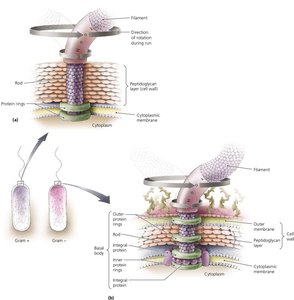
Flagella
Flagella are long, whip-like structures responsible for bacterial motility. Not all bacteria possess flagella.
Structure: Composed of filament, hook, and basal body; basal body anchors to cell wall.
Function: Rotation propels bacteria; movement can be clockwise or counterclockwise, resulting in 'runs' and 'tumbles'.
Arrangement: Flagella may be monotrichous, lophotrichous, amphitrichous, or peritrichous.


Fimbriae and Pili
Fimbriae and pili are surface appendages distinct from flagella, involved in adhesion and genetic exchange.
Fimbriae: Sticky, bristlelike projections; shorter than flagella; important in biofilm formation.
Pili: Special type of fimbriae; longer than fimbriae but shorter than flagella; involved in DNA transfer (conjugation).


Bacterial Cell Walls
Structure and Function
Bacterial cell walls provide structural support, shape, and protection from osmotic forces. They are composed primarily of peptidoglycan, a polymer of sugars and amino acids.
Peptidoglycan: Consists of N-acetylglucosamine (NAG) and N-acetylmuramic acid (NAM) linked by peptide crossbridges.
Shape: Cell walls give bacteria characteristic shapes (cocci, bacilli, etc.).
Antibiotic Target: Cell wall synthesis is targeted by antibiotics.


Gram-Positive vs. Gram-Negative Cell Walls
Bacterial cell walls are classified as Gram-positive or Gram-negative based on their structure and staining properties.
Gram-Positive: Thick peptidoglycan layer, teichoic acids, purple after Gram stain, may contain mycolic acid.
Gram-Negative: Thin peptidoglycan layer, outer membrane with lipopolysaccharide (LPS), pink after Gram stain; lipid A in LPS can cause toxic effects.
Feature | Gram-Positive | Gram-Negative |
|---|---|---|
Peptidoglycan | Thick | Thin |
Teichoic acids | Present | Absent |
Outer membrane | Absent | Present (with LPS) |
Gram stain color | Purple | Pink |
Mycolic acid | May be present | Absent |
Bacteria Without Cell Walls
Some bacteria, such as Mycoplasma, lack cell walls and are often mistaken for viruses due to their small size. However, they possess other prokaryotic features such as ribosomes.
Bacterial Cytoplasmic Membranes
Structure
The cytoplasmic membrane is a phospholipid bilayer containing integral and peripheral proteins. The fluid mosaic model describes its dynamic nature.
Phospholipid Bilayer: Provides selective permeability and structural integrity.
Proteins: Integral and peripheral proteins facilitate transport and communication.
Function
The membrane controls passage of substances, maintains gradients, and in photosynthetic bacteria, harvests light energy.
Selective Permeability: Only certain substances can cross.
Transport Mechanisms: Includes passive (diffusion, facilitated diffusion, osmosis) and active processes.
Electrical Gradient: Maintains membrane potential.
Passive Transport
Passive transport processes do not require energy and include diffusion, facilitated diffusion, and osmosis.
Diffusion: Movement of molecules from high to low concentration.
Facilitated Diffusion: Movement via protein channels.
Osmosis: Diffusion of water across a semipermeable membrane.
Example: Effects of isotonic, hypertonic, and hypotonic solutions on cells. 

Summary Table: Cell Structure and Function
Feature | Prokaryotes | Eukaryotes |
|---|---|---|
Nucleus | Absent | Present |
Organelles | Absent | Present |
Cell wall | Peptidoglycan (if present) | Cellulose/chitin (if present) |
Size | 1.0 µm or smaller | 10–100 µm |
Examples | Bacteria, Archaea | Algae, Protozoa, Fungi, Animals, Plants |
Additional info: Where original content was brief, academic context was added to clarify definitions, examples, and comparisons. All images included are directly relevant to the adjacent explanations and reinforce key concepts in cell structure and function.
