 Back
BackCell Structure and Function: Microbiology Study Notes
Study Guide - Smart Notes
 Tailored notes based on your materials, expanded with key definitions, examples, and context.
Cell Structure and Function
Characteristics of Life in Microbes
Microbes exhibit fundamental processes of life, which distinguish living organisms from non-living matter. These processes include growth, reproduction, responsiveness, and metabolism.
Growth: Increase in size or number of cells.
Reproduction: Ability to produce new cells or organisms.
Responsiveness: Reacting to environmental stimuli.
Metabolism: Chemical reactions that provide energy and build cellular structures.
Example: Mycoplasma, the smallest free-living microbe, is alive despite being nonmotile because it performs all other life processes.
Types of Cells: Prokaryotic vs. Eukaryotic
Cells are classified as prokaryotic or eukaryotic based on structural features. This distinction is fundamental in microbiology.
Prokaryotes:
Lack a nucleus
Simultaneously read DNA and synthesize proteins
Lack internal membrane-bound organelles
Typically 1.0 µm or smaller
Includes bacteria and archaea
Eukaryotes:
Have a nucleus
Contain internal membrane-bound organelles
Larger: 10–100 µm
More complex structure
Includes algae, protozoa, fungi, animals, and plants
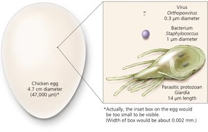
Cell Size Comparison
Microbial cells vary greatly in size, from viruses to protozoa and even multicellular organisms.
Viruses: ~0.3 µm diameter
Bacteria: ~1 µm diameter
Protozoa: ~14 µm length
Chicken egg: 47,000 µm diameter (for scale)
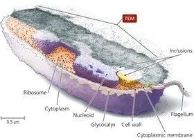
External Structures of Bacterial Cells
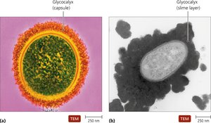
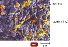
Glycocalyces
Bacterial cells may be surrounded by a glycocalyx, a gelatinous, sticky substance composed of polysaccharides, polypeptides, or both.
Capsule: Organized, firmly attached; protects against host recognition.
Slime layer: Loosely attached, water-soluble; aids in surface attachment.
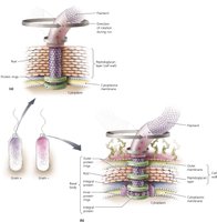
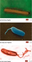
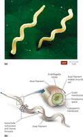
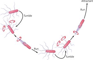
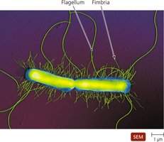
Flagella
Flagella are long, whip-like structures responsible for bacterial motility. Not all bacteria possess flagella.
Structure: Composed of filament, hook, and basal body. The basal body anchors the flagellum to the cell wall.
Function: Rotation propels bacteria; movement can be toward (runs) or away (tumbles) from stimuli (taxis).
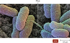
Fimbriae and Pili
Fimbriae and pili are surface structures that facilitate attachment and genetic exchange.
Fimbriae: Sticky, bristlelike projections; important in biofilm formation and adherence.
Pili: Longer than fimbriae, fewer per cell; specialized for DNA transfer (conjugation).
Bacterial Cell Walls
Structure and Function
Bacterial cell walls provide structural support, shape, and protection from osmotic forces. They are a target for antibiotics and contribute to characteristic cell shapes.
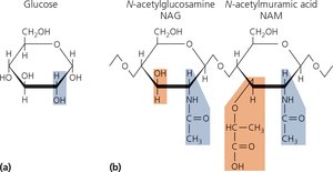
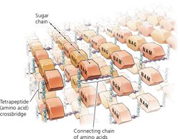
Peptidoglycan Composition
The main component of bacterial cell walls is peptidoglycan, a polymer of sugars and amino acids.
N-acetylglucosamine (NAG) and N-acetylmuramic acid (NAM) are the primary sugars.
Peptidoglycan forms a mesh-like structure with cross-linked chains.
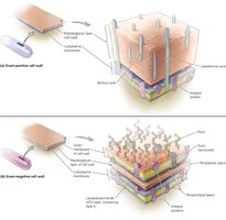
Gram-Positive vs. Gram-Negative Cell Walls
Bacteria are classified based on cell wall structure, which affects staining and susceptibility to antibiotics.
Gram-Positive:
Thick peptidoglycan layer
Contains teichoic acids and lipoteichoic acids
Stains purple in Gram stain
Acid-fast bacteria have mycolic acid for desiccation resistance
Gram-Negative:
Thin peptidoglycan layer
Outer membrane with phospholipids, proteins, and lipopolysaccharide (LPS)
Lipid A in LPS can cause fever and shock
Stains pink in Gram stain

Bacteria Without Cell Walls
Some bacteria lack cell walls and are often mistaken for viruses due to their small size. However, they possess other prokaryotic features such as ribosomes.
Bacterial Cytoplasmic Membranes
Structure
The cytoplasmic membrane is a phospholipid bilayer with embedded proteins, described by the fluid mosaic model.
Integral proteins: Span the membrane
Peripheral proteins: Attached to the membrane surface
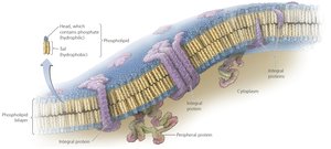
Function
The membrane controls passage of substances, maintains gradients, and in photosynthetic bacteria, harvests light energy. It is selectively permeable and proteins facilitate transport.
Cytoplasm of Bacteria
Cytosol and Inclusions
The cytosol is the liquid portion of the cytoplasm, containing water and DNA in the nucleoid region. Inclusions are reserve deposits of chemicals.
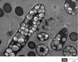
Endospores
Endospores are unique, highly resistant structures produced by some bacteria as a defense against unfavorable conditions. They can survive extreme heat, radiation, and chemicals.

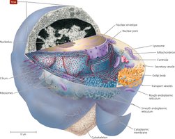
Cytoplasm of Eukaryotes
Nonmembranous Organelles: Ribosomes
Ribosomes are the sites of protein synthesis. Prokaryotic ribosomes are 70S, while eukaryotic ribosomes are 80S, reflecting structural differences.
Nonmembranous Organelles: Cytoskeleton
The cytoskeleton is composed of protein fibers and plays roles in cell division, shape, DNA segregation, and movement.
Summary Table: Gram-Positive vs. Gram-Negative Cell Walls
Feature | Gram-Positive | Gram-Negative |
|---|---|---|
Peptidoglycan Thickness | Thick | Thin |
Teichoic Acids | Present | Absent |
Outer Membrane | Absent | Present |
LPS (Lipid A) | Absent | Present |
Gram Stain Color | Purple | Pink |
Resistance to Desiccation | High (acid-fast) | Lower |
Key Equations
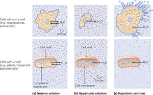
Osmosis: Movement of water across a selectively permeable membrane.
Peptidoglycan Structure: Repeating units of NAG and NAM linked by peptide bridges.
Conclusion
Understanding cell structure and function is fundamental in microbiology, providing insight into microbial classification, physiology, and the mechanisms underlying their survival and pathogenicity.
