 Back
BackCell Structure and Function: Microbiology Study Notes
Study Guide - Smart Notes
 Tailored notes based on your materials, expanded with key definitions, examples, and context.
Cell Structure and Function
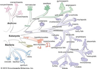
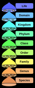
Domains of Life & Biological Classification
The classification of life forms is fundamental to microbiology, as it helps organize organisms based on shared characteristics. The three domains—Bacteria, Archaea, and Eukaryotes—are distinguished by cellular structure and genetic differences. Viruses, while not classified within these domains, are important in microbiology due to their unique properties.
Bacteria: Prokaryotic, single-celled organisms with diverse metabolic capabilities.
Archaea: Prokaryotic, often extremophiles, distinct from bacteria in cell wall composition and genetics.
Eukaryotes: Organisms with membrane-bound organelles, including plants, animals, fungi, and protists.
Viruses: Acellular entities, require host cells for replication.
Characteristics Common to All Life
All cellular life shares several fundamental characteristics, though viruses differ in key ways.
Growth: Increase in size; occurs in all cellular life, not in viruses.
Reproduction: Increase in number; all cells reproduce, viruses rely on host cells.
Responsiveness: Ability to react to environmental stimuli (e.g., taxis); present in all cells, limited in viruses.
Metabolism: Use of nutrients for energy; all cells metabolize, viruses use host metabolism.
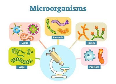
Types of Microorganisms
Microbiology studies a variety of microorganisms, each with unique structural and functional properties.
Bacteria
Archaea
Fungi
Protists
Viruses
Algae
Cell Structure: Prokaryotes vs. Eukaryotes
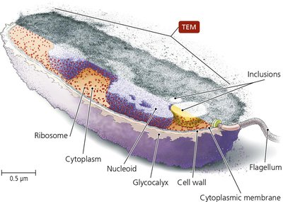
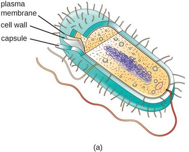
Prokaryotic Cell Structure
Prokaryotic cells (Bacteria and Archaea) lack membrane-bound organelles and a nucleus. Their cellular organization is simpler but highly efficient for survival in diverse environments.
Nucleoid: Region containing circular DNA.
Ribosomes: Sites of protein synthesis.
Cell wall: Provides structure and protection.
Glycocalyx: External layer for protection and adherence.
Flagella: Motility structures.
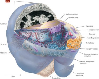
Eukaryotic Cell Structure
Eukaryotic cells possess membrane-bound organelles, including a nucleus, and are structurally more complex. This allows compartmentalization of functions and greater cellular specialization.
Nucleus: Contains linear DNA, control center.
Mitochondria: Site of aerobic ATP production.
Endoplasmic reticulum: Protein and lipid synthesis.
Golgi apparatus: Protein modification and sorting.
Lysosomes: Breakdown of nutrients and cellular debris.
Cytoskeleton: Structural support and movement.
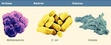
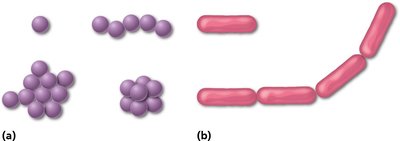
Comparison of Domains: Cell Morphology
Microbial domains can be visually distinguished by cell shape and structure.
Archaea: Often spherical or irregular (e.g., Methanosarcina).
Bacteria: Rod-shaped, spherical, or spiral (e.g., E. coli).
Eukarya: Variable shapes, often larger and more complex (e.g., Amoeba).
Organelles: Prokaryotes vs. Eukaryotes
Organelles are specialized structures within cells. Prokaryotes have fewer organelles, while eukaryotes possess both nonmembranous and membranous organelles.
Organelle | Function | Prokaryotes | Eukaryotes |
|---|---|---|---|
Ribosomes | Protein synthesis | Present in all | Present in all |
Cytoskeleton | Shape/support | Present in some | Present in all |
Centrosome/centrioles | Mitosis/cytokinesis | Absent | Present in animals |
Nucleus | Control center | Absent | Present in all |
Endoplasmic reticulum | Transport/lipid synthesis | Absent | Present in all |
Golgi apparatus | Secretion | Absent | Present in some |
Lysosomes | Breakdown | Absent | Present in some |
Peroxisomes | Neutralize toxins | Absent | Present in some |
Mitochondria | ATP production | Absent | Present in most |
Chloroplasts | Photosynthesis | Absent | Present in plants/algae |
External Structures of Cells
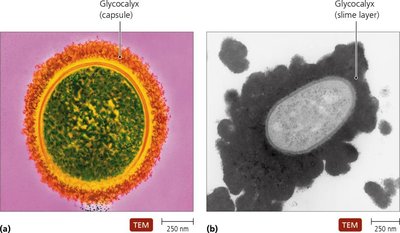
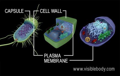
Glycocalyces
The glycocalyx is a gelatinous, sticky substance surrounding the outside of some cells, providing protection and aiding in adherence. In bacteria, it exists as either a capsule or a slime layer.
Capsule: Thick, tightly packed; protects from phagocytosis.
Slime layer: Loose, water-soluble; enhances adherence.
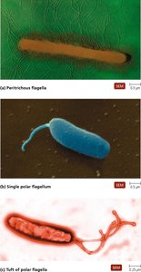
Flagella
Flagella are long, whip-like extensions used for motility. Their arrangement and structure vary among bacteria, archaea, and eukaryotes.
Structure: Composed of filament, hook, and basal body.
Protein: Made of flagellin in bacteria.
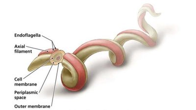
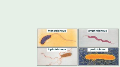
Arrangements: Peritrichous (cover cell), polar (at ends), endoflagella (spirochetes).
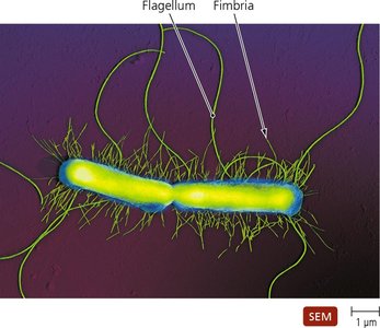
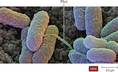
Fimbriae and Pili
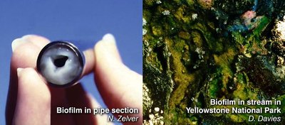
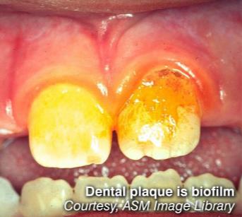
Fimbriae are short, bristle-like projections used for adherence and biofilm formation. Pili are longer and specialized for DNA transfer (conjugation).
Fimbriae: Important in biofilms, adherence to surfaces.
Pili: Transfer DNA between cells; not true sexual reproduction.
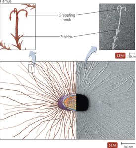
Archaeal External Structures
Archaea possess similar external structures to bacteria, including glycocalyces, flagella, and fimbriae. Unique to some archaea are hami, which are filamentous structures with hooks for attachment.
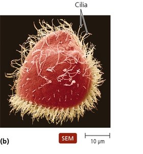
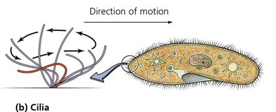
Eukaryotic External Structures
Eukaryotic cells may have glycocalyx, flagella (whip-like motion), and cilia (short, numerous, coordinated movement). Cilia are used for motility and moving substances across cell surfaces.
Comparison Summary Across Domains
Characteristic | Archaea | Bacteria | Eukaryotes |
|---|---|---|---|
Nucleus | Absent | Absent | Present |
Membrane-bound organelles | Absent | Present in few | Various types present |
Glycocalyx | Present | Capsule/slime layer | Present in some |
Flagella | Some, rotate | Some, rotate | Some, undulate |
Cilia | Absent | Absent | Present in some |
Fimbriae/Pili | Present in some | Present in some | Absent |
Hami | Present in some | Absent | Absent |
Cell wall | Most, no peptidoglycan | Most, peptidoglycan | Plants, algae, fungi |
Cytoplasmic membrane | All | All | All |
Cytosol | All | All | All |
Endospores | Absent | Some | Absent |
Chromosomes | Single, circular | Single, circular | Linear, multiple |
Cell Walls & Cell/Plasma Membranes
Bacterial Cell Walls
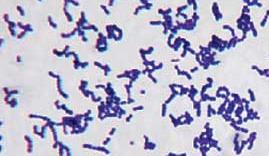
Bacterial cell walls provide structure, shape, and protection. The main shapes are cocci (spherical) and bacilli (rod-shaped). Cell walls are composed of peptidoglycan and are classified as Gram-positive or Gram-negative based on staining properties.
Gram-positive: Thick peptidoglycan, teichoic acids, purple stain.
Gram-negative: Thin peptidoglycan, outer membrane with LPS, pink stain.
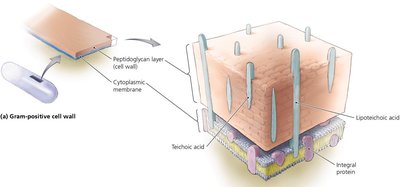
Gram-Positive Bacteria Cell Wall
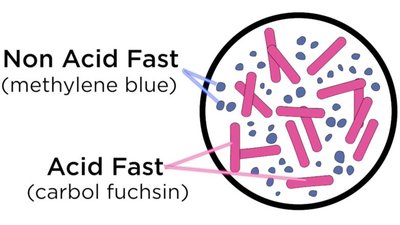
Gram-positive bacteria have a thick peptidoglycan layer, making them more susceptible to antibiotics. Some possess mycolic acid, requiring acid-fast staining.
Teichoic and lipoteichoic acids: Anchor cell wall to membrane.
Acid-fast bacteria: Retain red/pink stain due to mycolic acid (e.g., Mycobacterium).
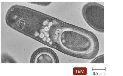
Gram-Positive Bacteria: Endospores
Many Gram-positive bacteria can form endospores, which protect DNA and allow survival in harsh conditions. Endospores are mainly found in Bacillus and Clostridium genera.
Gram-Negative Bacteria Cell Wall
Gram-negative bacteria have a thin peptidoglycan layer and two plasma membranes. The outer membrane contains lipopolysaccharide (LPS), with Lipid A contributing to pathogenic effects.
Lipid A: Can cause fever, shock, and blood clotting when released.
Gram stain: Pink due to thin peptidoglycan.
Gram + vs. Gram -: Side by Side Comparison
Gram-positive and Gram-negative bacteria differ in cell wall structure, staining, and susceptibility to antibiotics.
Feature | Gram-Positive | Gram-Negative |
|---|---|---|
Peptidoglycan | Thick | Thin |
Outer membrane | Absent | Present |
Teichoic acids | Present | Absent |
LPS | Absent | Present |
Stain | Purple | Pink |
Antibiotic susceptibility | Higher | Lower |
Membrane Structure & Function
Membrane Structure and Functions
The cell membrane controls passage of substances, maintains gradients, and is selectively permeable. Proteins facilitate transport across the membrane.
Selective permeability: Only certain substances can cross.
Transport proteins: Allow movement of ions and molecules.
Concentration/electrical gradients: Essential for cell function.
Membrane Permeability and Transport
Transport across membranes occurs via passive (no energy) or active (requires energy) mechanisms.
Passive transport: Simple diffusion, facilitated diffusion, osmosis.
Active transport: Requires ATP to move substances against gradients.
Osmosis
Osmosis is the diffusion of water across a semipermeable membrane. The effects of isotonic, hypertonic, and hypotonic solutions on cells are important in microbiology.
Isotonic: No net water movement.
Hypertonic: Water leaves cell, causing shrinkage.
Hypotonic: Water enters cell, causing swelling.
Equations
Diffusion and osmosis can be described mathematically:
Fick's Law of Diffusion:
Osmotic Pressure:
Where: J = flux, D = diffusion coefficient, dC/dx = concentration gradient, \Pi = osmotic pressure, i = van't Hoff factor, M = molarity, R = gas constant, T = temperature.
*Additional info: Equations and mathematical context added for completeness.*
