 Back
Cell Structure and Function: Prokaryotes vs. Eukaryotes
Study Guide - Smart Notes
 Tailored notes based on your materials, expanded with key definitions, examples, and context.
Cell Structure: Prokaryotes vs. Eukaryotes
Overview of Cell Types
Prokaryotic and eukaryotic cells represent two fundamental organizational types in microbiology. Understanding their structural differences is essential for grasping microbial physiology and classification.
Prokaryotes: Include bacteria and archaea; lack membrane-bound organelles.
Eukaryotes: Include fungi, protozoa, algae, plants, and animals; possess membrane-bound organelles.
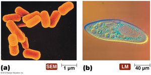
External Structures of Prokaryotic Cells
Prokaryotic cells possess several external structures that contribute to their survival and pathogenicity.
Flagella: Long, whip-like appendages used for locomotion. Arrangements include monotrichous, lophotrichous, amphitrichous, and peritrichous.
Pili: Rigid, hollow tubes for attachment and DNA transfer.
Fimbriae: Short, hair-like fibers for attachment, especially in biofilms.
Glycocalyx: Includes slime layer (loose, protective, aids in attachment) and capsule (firmly attached, protects from phagocytosis).
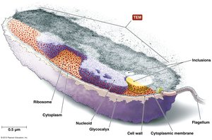
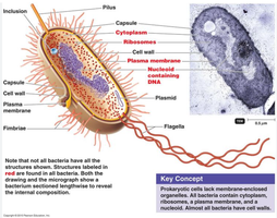
Prokaryotic Cell Envelope and Wall
The cell envelope consists of the cell wall, plasma membrane, and, in some cases, an outer membrane.
Cell Wall: Provides shape, structural support, and protection from lysis. Composed mainly of peptidoglycan.
Gram-Positive: Thick peptidoglycan layer, teichoic acids, stains purple.
Gram-Negative: Thin peptidoglycan layer, outer membrane with lipopolysaccharide (LPS), stains pink.
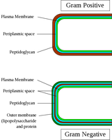
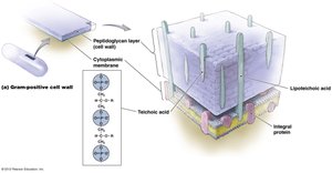
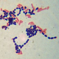
Table: Comparative Characteristics of Gram-Positive and Gram-Negative Bacteria
Characteristic | Gram-Positive | Gram-Negative |
|---|---|---|
Gram Reaction | Retains crystal violet, stains blue/purple | Can be decolorized, stains red/pink |
Peptidoglycan Layer | Thick, multilayered | Thin, single-layered |
Teichoic Acid | Present | Absent |
Outer Membrane | Absent | Present |
LPS Content | Low | High |
Flagellar Structure | 2 rings in basal body | 4 rings in basal body |
Internal Structures of Prokaryotic Cells
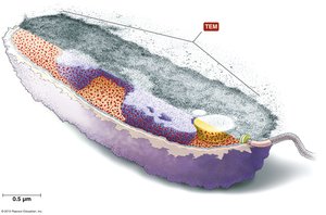
Prokaryotic cells contain several internal components essential for their function.
Cytoplasm: Gelatinous solution, site of biochemical reactions.
Nucleoid: Region containing a single, circular DNA molecule.
Ribosomes: 70S type, site of protein synthesis.
Inclusions: Reserve deposits of chemicals.
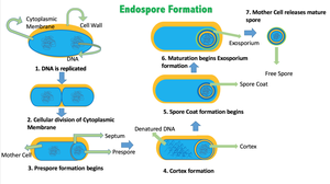
Endospores: Highly resistant structures formed by some bacteria for survival under harsh conditions.
Plasmids: Small, circular DNA molecules conferring traits like drug resistance.
Cytoskeleton: Protein fibers involved in cell shape and division.
Eukaryotic Cell Structure
External Structures of Eukaryotic Cells
Eukaryotic cells have more complex external structures.
Glycocalyces: Less organized than prokaryotic capsules; function in cell recognition and protection.
Cell Wall: Present in fungi, algae, plants, and some protozoa; absent in animal cells.
Eukaryotic Cytoplasmic Membrane
Fluid Mosaic Model: Composed of phospholipids and proteins; contains steroid lipids for fluidity.
Membrane Rafts: Regions of lipids and proteins for signaling and transport.
Internal Structures of Eukaryotic Cells
Eukaryotic cells contain numerous membrane-bound organelles.
Nucleus: Contains most of the cell’s DNA; surrounded by nuclear envelope.
Endoplasmic Reticulum (ER): Rough ER (with ribosomes) and Smooth ER (without ribosomes); functions in protein and lipid synthesis.
Golgi Body: Processes and packages molecules for export.
Lysosomes, Peroxisomes, Vacuoles: Store and transfer chemicals; lysosomes digest macromolecules, peroxisomes degrade toxins.
Mitochondria: Site of ATP production; contains its own DNA and 70S ribosomes.
Chloroplasts: Found in photosynthetic eukaryotes; site of photosynthesis.
Cytoskeleton: Network of fibers for cell shape, movement, and organelle anchoring.
Ribosomes: 80S type, larger than prokaryotic ribosomes.
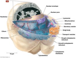
Comparative Summary: Prokaryotes vs. Eukaryotes
Key Differences
Prokaryotes: No membrane-bound organelles, nucleoid region, 70S ribosomes, cell wall with peptidoglycan.
Eukaryotes: Membrane-bound organelles, nucleus, 80S ribosomes, cell wall (if present) lacks peptidoglycan.
Endosymbiotic Theory
The endosymbiotic theory explains the origin of mitochondria and chloroplasts in eukaryotic cells.
Small aerobic prokaryotes became internal symbionts within larger anaerobic prokaryotes.
These symbionts evolved into mitochondria and chloroplasts.
Take Home Points
Know the major prokaryotic structures and their functions.
Know the major eukaryotic structures and their functions.
Be able to compare and contrast structures between eukaryotic and prokaryotic cells.
Additional info:
Prokaryotic cell walls are a primary target for antibiotics, as disruption leads to cell lysis.
Gram-negative bacteria are generally more resistant to antimicrobial agents due to their outer membrane.
Endospores are highly resistant to heat, desiccation, chemicals, and radiation.
