 Back
BackCell Structure and Function: Prokaryotic and Eukaryotic Cells
Study Guide - Smart Notes
 Tailored notes based on your materials, expanded with key definitions, examples, and context.
Cell Structure and Function
Overview of Cellular Life
All living things share four fundamental processes: growth, reproduction, responsiveness, and metabolism. These processes distinguish living cells from nonliving matter and are essential for life.
Growth: Increase in size.
Reproduction: Increase in number, either asexually or sexually.
Responsiveness: Ability to respond to environmental stimuli (taxis).
Metabolism: Controlled chemical reactions to obtain energy and build cellular structures. Energy is stored in adenosine triphosphate (ATP).
Comparison of Prokaryotic and Eukaryotic Cells
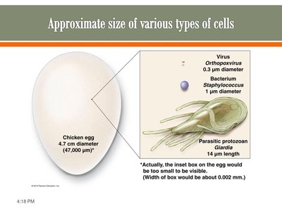
General Features and Size
Cells are classified as prokaryotic or eukaryotic based on structural differences, particularly the presence or absence of a nucleus and membrane-bound organelles.
Prokaryotes: Simple structure, small size (1–10 µm), no nucleus, no membrane-bound organelles. Includes Bacteria and Archaea.
Eukaryotes: Complex structure, larger size (10–100 µm), nucleus present, membrane-bound organelles. Includes algae, protozoa, fungi, animals, and plants.
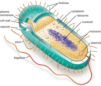
External Structures of Prokaryotic Cells
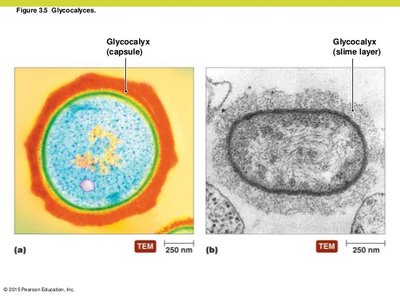
Capsule and Glycocalyx
Many prokaryotic cells possess external structures that aid in protection, adherence, and evasion of host defenses.
Capsule: Firmly attached, composed of polysaccharides or proteins. Protects bacteria from host immune recognition.
Glycocalyx: General term for external polysaccharide or polypeptide layer; includes capsules and slime layers.
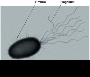
Flagella, Fimbriae, and Pili
These structures facilitate movement, adherence, and genetic exchange.
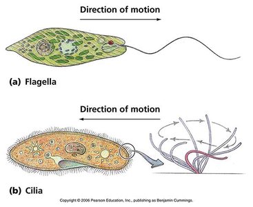
Flagella: Long, whip-like structures for motility. Prokaryotic flagella rotate; eukaryotic flagella undulate.
Fimbriae: Short, bristle-like fibers for adherence to surfaces and biofilm formation.
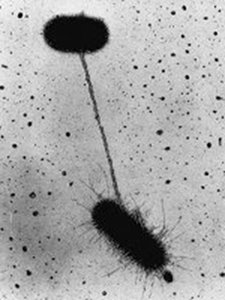
Pili: Longer than fimbriae, used for DNA transfer (conjugation).
Prokaryotic Cell Walls
Peptidoglycan Structure
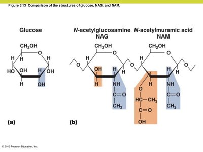
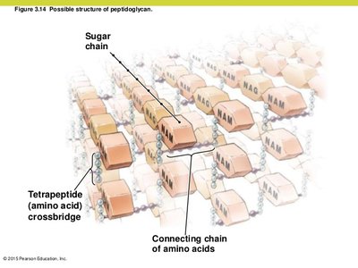
The cell wall provides structural support and protection against osmotic forces. Most prokaryotic cell walls are composed of peptidoglycan, a mesh-like polymer of sugars and amino acids.
Peptidoglycan: Composed of alternating N-acetylglucosamine (NAG) and N-acetylmuramic acid (NAM) sugars, cross-linked by peptide bridges.
Shapes: Genetically determined; includes bacillus (rod-shaped) and coccus (spherical).
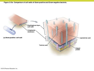
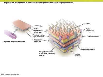
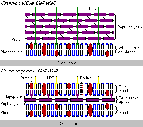
Gram-Positive vs. Gram-Negative Cell Walls
Bacterial cell walls are classified as Gram-positive or Gram-negative based on their structure and staining properties.
Gram-Positive: Thick peptidoglycan layer, contains teichoic acids, stains purple. Retains crystal violet dye.
Gram-Negative: Thin peptidoglycan layer, outer membrane with lipopolysaccharide (LPS), stains pink. Contains endotoxin (Lipid A).
Bacterial Cytoplasmic Membrane
Structure and Function
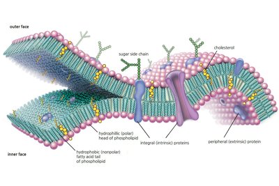
The cytoplasmic membrane is a phospholipid bilayer with embedded proteins, described by the fluid mosaic model. It is selectively permeable and maintains concentration and electrical gradients.
Phospholipid bilayer: Hydrophilic heads face outward; hydrophobic tails face inward.
Functions: Selective permeability, energy harvesting (in photosynthetic bacteria), transport of molecules, maintenance of gradients.
Osmosis and Effects of Solutions
Osmosis is the movement of water across a semipermeable membrane. The effects of isotonic, hypertonic, and hypotonic solutions differ among animal, plant, and bacterial cells.
Isotonic: No net movement of water.
Hypertonic: Water leaves the cell; plasmolysis may occur.
Hypotonic: Water enters the cell; may cause lysis in animal cells.
Internal Structures of Prokaryotic Cells
Cytoplasm and Ribosomes
The cytoplasm contains water, ions, enzymes, and the nucleoid region with DNA. Ribosomes (70S) are nonmembranous organelles responsible for protein synthesis.
Nucleoid: Region containing the bacterial chromosome.
Inclusions: Storage deposits of molecules.
Ribosomes: Translate mRNA into proteins.
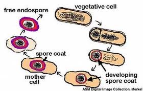
Endospores
Endospores are highly resistant structures formed by some bacteria as a defense against unfavorable conditions. They are not reproductive structures but allow survival in extreme environments.
Formation: Vegetative cell transforms into an endospore when nutrients are limited.
Resistance: Heat, radiation, chemicals.
Example: Bacillus anthracis forms endospores, relevant in bioterrorism.
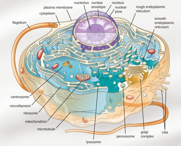
Eukaryotic Cell Structures
External Structures
Eukaryotic cells may have glycocalyces, cell walls, flagella, and cilia. Glycocalyces are less organized than prokaryotic capsules and aid in cell recognition and protection.
Cell Walls: Composed of polysaccharides (cellulose in plants, chitin in fungi).
Flagella: Covered by cell membrane, contain microtubules, undulate for movement.
Cilia: Short, numerous, used for locomotion and moving substances past cell surface.
Membranous Organelles
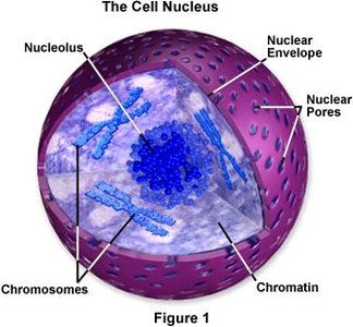
Nucleus: Contains DNA as chromatin, surrounded by nuclear envelope.
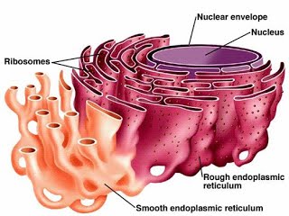
Endoplasmic Reticulum (ER): Smooth ER synthesizes lipids; rough ER has ribosomes for protein synthesis.
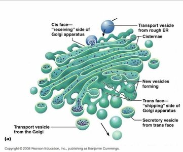
Golgi Body: Processes and packages molecules for export.
Lysosomes: Contain enzymes for digestion of cellular components.
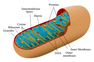
Mitochondria: Site of ATP production; contains 70S ribosomes and circular DNA.
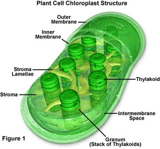
Chloroplasts: Light-harvesting organelles in photosynthetic eukaryotes; contain 70S ribosomes and circular DNA.
Endosymbiotic Theory
Origin of Eukaryotic Organelles
The endosymbiotic theory, proposed by Dr. Lynn Margulis, explains the origin of mitochondria and chloroplasts as formerly free-living prokaryotes engulfed by ancestral eukaryotic cells.
Evidence: Both organelles have double membranes, circular DNA, and 70S ribosomes similar to prokaryotes.
Key Scientists in Microbiology
Hans Christian Gram: Invented the Gram stain method for bacterial classification.
Lynn Margulis: Proposed the endosymbiotic theory.
Bruce Ivins: Associated with anthrax research and bioterrorism investigations.
Summary Table: Comparison of Cell Types
Feature | Prokaryotes | Eukaryotes |
|---|---|---|
Nucleus | No | Yes |
Membrane-bound organelles | No | Yes |
Cell wall composition | Peptidoglycan (Bacteria) | Polysaccharides (Cellulose, Chitin) |
Ribosome size | 70S | 80S (except mitochondria/chloroplasts: 70S) |
Flagella structure | Rotating, not covered by membrane | Undulating, covered by membrane |
Additional info: This study guide covers all major aspects of cell structure and function relevant to microbiology, including comparisons, structural details, and key processes. It is suitable for exam preparation and foundational understanding.
