 Back
BackCell Structure and Function: Prokaryotic and Eukaryotic Microorganisms
Study Guide - Smart Notes
 Tailored notes based on your materials, expanded with key definitions, examples, and context.
Cell Structure and Function
Types of Cellular Microorganisms
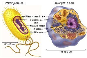
Microorganisms are classified into two main cellular types: prokaryotic and eukaryotic. Understanding their structural differences is fundamental in microbiology.
Prokaryotic cells: Lack a true nucleus and membrane-bound organelles. Examples include bacteria and archaea.
Eukaryotic cells: Possess a nucleus and various membrane-bound organelles. Examples include fungi, protozoa, algae, and animal cells.
Acellular Microorganisms: Viruses
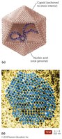
Viruses are considered acellular and non-living. They are obligate intracellular parasites, meaning they require a host cell to replicate. Viruses exist in two states:
Extracellular state (virion): Composed of nucleic acid, a protein capsid, and sometimes an envelope.
Intracellular state: Capsid/envelope is removed, and the virus exists as nucleic acid within the host cell.
Bacterial Cell Morphology
Size, Shape, and Arrangement
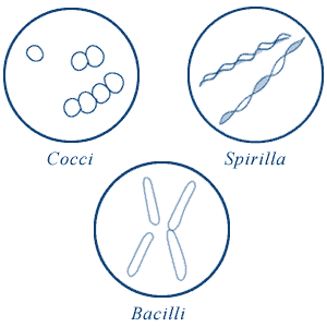
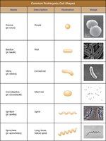
Bacterial cells exhibit a variety of shapes and arrangements, which are important for identification and classification.
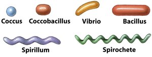
Basic shapes: Bacillus (rod), coccus (spherical), spirillum (spiral).
Variations: Coccobacillus (short rod), vibrio (curved rod), spirochete (long, flexible spiral).
Name | Description | Illustration | Image |
|---|---|---|---|
Coccus | Round | Single sphere | Image of cocci |
Bacillus | Rod | Single rod | Image of bacilli |
Vibrio | Curved rod | Comma-shaped | Image of vibrio |
Coccobacillus | Short rod | Oval | Image of coccobacillus |
Spirillum | Spiral | Rigid spiral | Image of spirillum |
Spirochete | Long, flexible spiral | Flexible spiral | Image of spirochete |
Bacterial Cell Arrangements
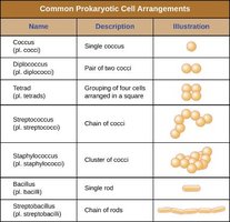
Bacteria can be arranged in characteristic patterns depending on their division and grouping.
Cocci: Single, pairs (diplococci), chains (streptococci), clusters (staphylococci), tetrads, sarcinae.
Bacilli: Single, pairs (diplobacilli), chains (streptobacilli), palisades.
Name | Description | Illustration |
|---|---|---|
Coccus | Single coccus | Single sphere |
Diplococcus | Pair of two cocci | Two spheres |
Tetrad | Grouping of four cells arranged in a square | Four spheres |
Streptococcus | Chain of cocci | Chain of spheres |
Staphylococcus | Cluster of cocci | Cluster of spheres |
Bacillus | Single rod | Single rod |
Streptobacillus | Chain of rods | Chain of rods |
Prokaryotic Cell Structure
Basic Prokaryotic Cell Structure
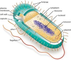
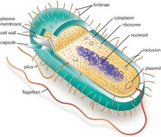
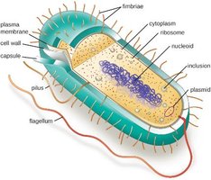
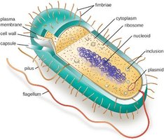
A typical prokaryotic cell contains a cell membrane, chromosomal DNA concentrated in a nucleoid, ribosomes, and a cell wall. Some prokaryotic cells may also possess flagella, pili, fimbriae, and capsules.
Cell Wall
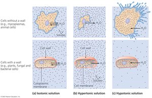
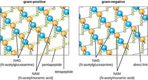
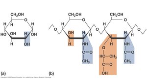
The cell wall is a critical structure in bacteria, providing shape and protection against osmotic lysis. The main component is peptidoglycan, a polymer of alternating N-acetylmuramic acid (NAM) and N-acetylglucosamine (NAG) subunits, cross-linked by peptide bridges.
Function: Maintains cell shape, prevents hypotonic lysis.
Structure: Peptidoglycan provides tensile strength.
Gram-Positive vs Gram-Negative Cell Walls
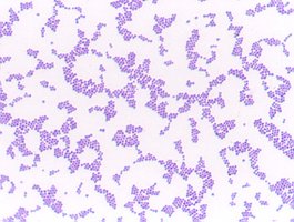
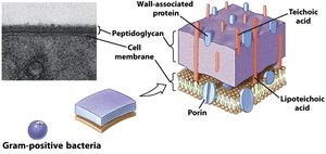
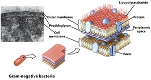
Bacteria are classified as Gram-positive or Gram-negative based on cell wall structure, which affects staining and antibiotic susceptibility.
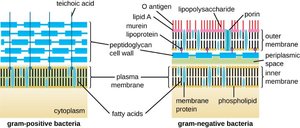
Gram-positive: Thick peptidoglycan layer (20–80 nm), teichoic acids, stains purple.
Gram-negative: Thin peptidoglycan layer (1–3 nm), outer membrane with lipopolysaccharide (LPS), periplasmic space, stains pink.
Cell Wall Variations: Mycobacteria and Mycoplasma
Some bacteria have unique cell wall structures or lack a cell wall entirely.
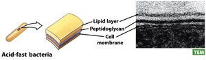
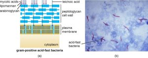
Mycobacteria: Possess a waxy mycolic acid layer, detected by acid-fast staining. Example: Mycobacterium tuberculosis.
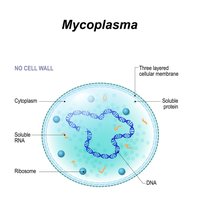
Mycoplasma: Lack a cell wall, resulting in variable shape. Example: Mycoplasma pneumoniae.
Cell Membrane Structure
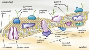
The bacterial cell membrane is a phospholipid bilayer with embedded proteins, glycoproteins, and glycolipids. It regulates transport, energy production, and cell signaling.
Internal Structures
Prokaryotic cells contain several internal structures essential for their function:
Cytoplasm: Gel-like substance where metabolic reactions occur.
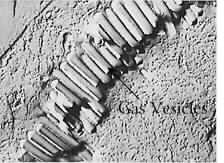
Inclusions: Storage granules for nutrients and other substances.
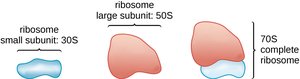
Ribosomes: Sites of protein synthesis (70S, composed of 30S and 50S subunits).
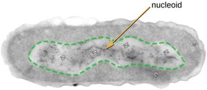
Nucleoid: Region containing the bacterial chromosome (usually a single, circular DNA molecule).
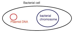
Plasmids: Small, extrachromosomal DNA molecules carrying non-essential genes, often for antibiotic resistance or virulence.
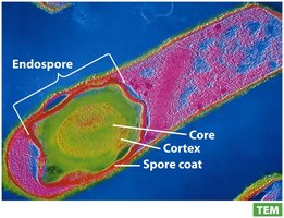
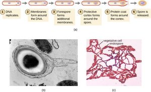
Endospores: Dormant, highly resistant structures formed by some genera (e.g., Bacillus, Clostridium).

External Structures
Flagella
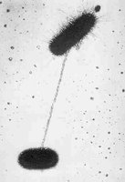
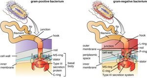
Flagella are whip-like appendages used for motility. Their structure consists of a basal body, hook, and filament. Flagella arrangements include monotrichous, amphitrichous, lophotrichous, and peritrichous.

Flagellar Movement: Runs vs Tumbles
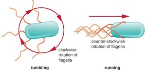
Bacterial movement is characterized by alternating runs (straight movement) and tumbles (random changes in direction), controlled by flagellar rotation.
Chemotaxis
Chemotaxis is the movement of bacteria toward or away from chemical stimuli. Positive chemotaxis is movement toward attractants, while negative chemotaxis is movement away from repellents.

Pili and Fimbriae
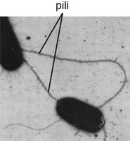
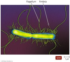
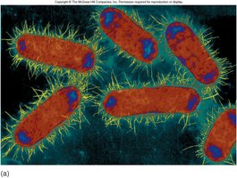
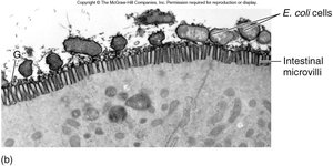
Pili are hair-like appendages used for attachment and conjugation (transfer of plasmid DNA). Fimbriae are shorter and more numerous, aiding in attachment to surfaces and other cells.
Glycocalyx: Capsules and Slime Layers
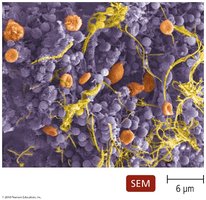
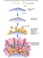
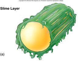
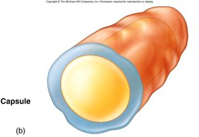
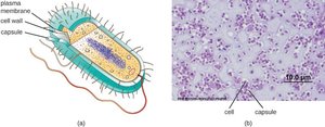
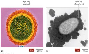
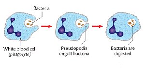
The glycocalyx is an external layer composed of polysaccharides. It can be organized as a capsule or unorganized as a slime layer. Functions include protection from drying out, prevention of phagocytosis, and aiding in attachment and biofilm formation.
Capsule: Organized, tightly bound, enhances virulence.
Slime layer: Loosely organized, aids in environmental survival.
Eukaryotic Cell Structure
General Eukaryotic Cell Structure
Eukaryotic cells are more complex, containing a nucleus and various organelles. External structures include glycocalyces (in animal and protozoan cells) and cell walls (in fungi and some protozoa).
External Eukaryotic Structures
Glycocalyces: Found in animal and most protozoan cells, absent in cells with cell walls.
Cell walls: Found in fungi and some protozoa, absent in animal cells.
Cell Membranes in Eukaryotes
Eukaryotic cell membranes are similar to those in bacteria but contain steroids (cholesterol in animal cells, ergosterol in fungi) for structural support.
Flagella and Cilia in Eukaryotes
Some eukaryotic cells possess flagella or cilia for movement. These structures are more complex than their prokaryotic counterparts.
Nonmembranous and Membranous Organelles
Eukaryotic cells contain both nonmembranous (e.g., ribosomes) and membranous organelles (e.g., mitochondria, endoplasmic reticulum). Eukaryotic ribosomes are larger (80S, composed of 60S and 40S subunits) and are not targeted by antibiotics that affect prokaryotic ribosomes.
