 Back
Cell Structure and Function: Prokaryotic and Eukaryotic Cells
Study Guide - Smart Notes
 Tailored notes based on your materials, expanded with key definitions, examples, and context.
Cell Walls and the Gram Stain Mechanism
Gram-Positive vs. Gram-Negative Bacteria
The Gram stain is a differential staining technique that distinguishes bacteria based on the structure of their cell walls. The outcome of the Gram stain is determined by the ability of the cell wall to retain the crystal violet-iodine complex during the decolorization step.
Gram-Positive Bacteria: Thick peptidoglycan layer retains the crystal violet-iodine complex, resulting in a purple color after staining.
Gram-Negative Bacteria: Thin peptidoglycan layer and an outer membrane; alcohol dissolves the outer membrane and washes out the crystal violet-iodine complex, so cells appear colorless until counterstained with safranin (red/pink).
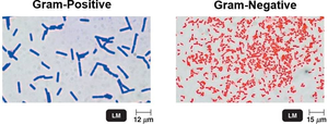
Key Steps in Gram Staining:
Application of crystal violet (primary stain)
Addition of iodine (mordant)
Alcohol wash (decolorization)
Counterstain with safranin
Mechanism: In Gram-positive cells, alcohol dehydrates the thick peptidoglycan, trapping the dye. In Gram-negative cells, alcohol disrupts the outer membrane and creates holes in the thin peptidoglycan, allowing the dye to escape.
Comparative Characteristics of Gram-Positive and Gram-Negative Bacteria
Feature | Gram-Positive | Gram-Negative |
|---|---|---|
Peptidoglycan Layer | Thick | Thin |
Outer Membrane | Absent | Present |
Teichoic Acids | Present | Absent |
Flagellar Basal Body | Two rings | Four rings |
Toxins Produced | Exotoxins | Endotoxins and exotoxins |
Penicillin Susceptibility | High | Low |
Lysozyme Sensitivity | High | Low |
Atypical Cell Walls
Acid-Fast Cell Walls
Some bacteria, such as Mycobacterium and Nocardia, have cell walls similar to Gram-positive bacteria but contain a waxy lipid (mycolic acid) bound to peptidoglycan. These cells retain carbolfuchsin dye even after acid-alcohol wash, making them acid-fast.
Acid-fast stain: Used to identify acid-fast organisms.
Mycoplasmas and Archaea
Mycoplasmas: Lack cell walls; sterols in the plasma membrane provide stability and resistance to lysis.
Archaea: May lack cell walls or have walls made of pseudomurein (lacking NAM and D-amino acids).
Structures Internal to the Cell Wall
The Plasma (Cytoplasmic) Membrane: Structure
The plasma membrane is a phospholipid bilayer with embedded proteins, enclosing the cytoplasm. It is essential for maintaining cellular integrity and mediating transport.
Phospholipid bilayer: Composed of hydrophilic heads and hydrophobic tails.
Proteins: Peripheral (surface) and integral (embedded) proteins; some are glycoproteins or glycolipids.
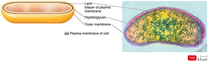
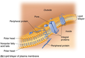
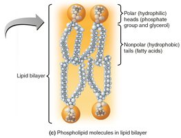
The Plasma (Cytoplasmic) Membrane: Function
Selective permeability: Allows passage of some molecules but not others.
Transport mechanisms: Simple diffusion, facilitated diffusion, osmosis, active transport, and group translocation.
Destruction by Antimicrobial Agents: Alcohols, quaternary ammonium compounds, and antibiotics like polymyxin can damage the plasma membrane, causing leakage of cell contents.
Cytoplasm
The cytoplasm is a thick, aqueous, and elastic substance inside the plasma membrane, containing water, proteins, carbohydrates, lipids, ions, DNA, ribosomes, and inclusions. The cytoskeleton provides structure, aids in cell division, and supports DNA movement.
The Nucleoid
Bacterial chromosome: Circular, double-stranded DNA containing genetic information, not enclosed by a nuclear envelope.
Plasmids: Small, extrachromosomal DNA circles carrying non-essential genes (e.g., antibiotic resistance), replicating independently and transferable between cells.
Ribosomes
Function: Sites of protein synthesis.
Structure: Composed of protein and rRNA; prokaryotic ribosomes are 70S (50S + 30S subunits).
Antibiotic targets: Streptomycin, gentamicin, erythromycin, and chloramphenicol inhibit prokaryotic ribosomes.
Inclusions
Inclusions are reserve deposits of nutrients and other substances within the cytoplasm.
Metachromatic granules (volutin): Phosphate reserves.
Polysaccharide granules, lipid inclusions, sulfur granules: Energy reserves.
Carboxysomes: Contain RuBisCO enzyme for CO2 fixation during photosynthesis.
Gas vacuoles: Protein-covered cylinders for buoyancy.
Magnetosomes: Iron oxide inclusions that help orient bacteria to magnetic fields.
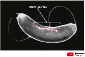
Endospores
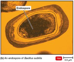
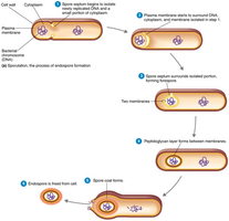
Endospores are highly resistant, dormant structures formed by certain bacteria (e.g., Bacillus and Clostridium) when nutrients are scarce. They can survive extreme conditions and are important in food safety.
Sporulation: Process of endospore formation.
Germination: Return of endospore to vegetative state.
Eukaryotic Cell Structures
Overview of Eukaryotic Cell Structures
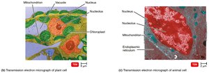
Eukaryotic cells contain membrane-bound organelles and a defined nucleus. Structures include the nucleus, mitochondria, chloroplasts, endoplasmic reticulum, Golgi complex, lysosomes, peroxisomes, and cytoskeleton.
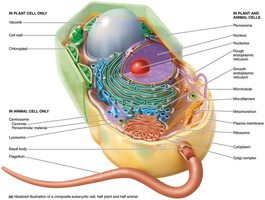
Flagella and Cilia
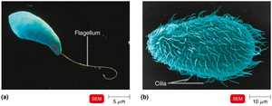
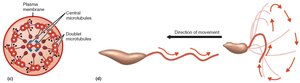
Flagella and cilia are projections used for locomotion or moving substances along the cell surface. Eukaryotic flagella are long and few, while cilia are short and numerous. Both are composed of microtubules made of tubulin and move in a wavelike manner.
Cell Wall and Glycocalyx
Cell wall: Found in plants, algae, and fungi; composed of carbohydrates (cellulose, chitin, glucan, mannan).
Glycocalyx: Carbohydrates bonded to proteins and lipids in the plasma membrane; found in animal cells, strengthens the cell surface, aids in attachment, and cell–cell recognition.
The Plasma (Cytoplasmic) Membrane in Eukaryotes
Structure: Similar to prokaryotes (phospholipid bilayer, proteins), but contains sterols and carbohydrates for attachment and recognition.
Function: Selective permeability, transport (simple/facilitated diffusion, osmosis, active transport), and endocytosis (phagocytosis, pinocytosis, receptor-mediated).
Cytoplasm and Cytoskeleton
Cytoplasm: Substance inside the plasma membrane and outside the nucleus.
Cytoskeleton: Microfilaments, intermediate filaments, and microtubules provide shape and support.
Ribosomes
Function: Sites of protein synthesis.
Structure: Eukaryotic ribosomes are 80S (60S + 40S subunits); some are membrane-bound (attached to ER), others are free in the cytoplasm.
Antibiotic sensitivity: Some antibiotics (e.g., erythromycin) target the 50S subunit, affecting prokaryotic but not eukaryotic ribosomes.
Organelles
Nucleus: Double membrane (nuclear envelope) enclosing DNA complexed with histones (chromatin); condenses into chromosomes during cell division.
Lysosomes: Vesicles from the Golgi complex containing digestive enzymes.
Mitochondria: Double membrane, site of ATP production (cellular respiration), contains 70S ribosomes and circular DNA, can reproduce independently.
Chloroplasts: Site of photosynthesis, contains 70S ribosomes and circular DNA.
The Evolution of Eukaryotes
Endosymbiotic Theory
The endosymbiotic theory explains the origin of eukaryotic cells. It proposes that larger bacterial cells engulfed smaller ones, which became organelles such as mitochondria and chloroplasts. The nucleus formed from infoldings of the plasma membrane.
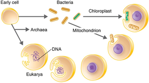
Evidence: Mitochondria and chloroplasts resemble bacteria in size and shape, have circular DNA, reproduce independently, possess 70S ribosomes, and have double membranes.
