 Back
Central Dogma, DNA Replication, Transcription, Translation, and Gene Regulation in Microbiology
Study Guide - Smart Notes
 Tailored notes based on your materials, expanded with key definitions, examples, and context.
Central Dogma of Molecular Biology
Overview of Information Flow
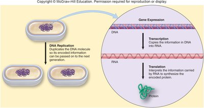
The central dogma of molecular biology describes the directional flow of genetic information within a cell: DNA is transcribed into RNA, which is then translated into protein. This process is fundamental to all living organisms and underpins cellular function and inheritance.
DNA Replication: Duplicates the DNA molecule so encoded information can be passed to the next generation.
Transcription: Copies information in DNA into RNA.
Translation: Interprets the information carried by RNA to synthesize the encoded protein.
DNA: Structure and Function
DNA as the Blueprint of Life
DNA is the hereditary material in all living cells, encoding the instructions for life. Its structure allows for the storage and transmission of vast amounts of information.
Nucleotides: DNA is composed of four nucleotides: adenine (A), thymine (T), cytosine (C), and guanine (G).
Genome: The complete set of genetic information, including chromosomes and plasmids.
Gene: The functional unit of DNA, encoding a gene product (usually a protein).
Genomics: The study of nucleotide sequences and their functions.
DNA Structure
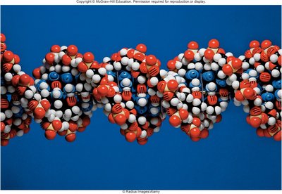
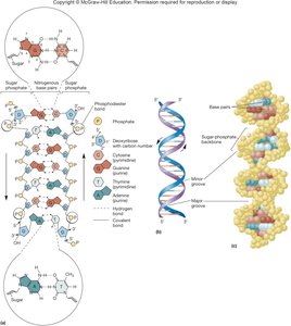
DNA is a double-stranded helix with specific base pairing and structural features that facilitate replication and gene expression.
Double Helix: Two strands twist around each other, forming major and minor grooves.
Base Pairing: Adenine pairs with thymine via two hydrogen bonds; guanine pairs with cytosine via three hydrogen bonds.
Phosphodiester Bonds: Link nucleotides together in each strand.
RNA Structure and Types
RNA Structure
RNA is a polymer of nucleotides similar to DNA but with key differences. Most RNA molecules are single-stranded, though some viral RNAs are double-stranded.
Bases: Adenine, guanine, cytosine, and uracil (instead of thymine).
Sugar: Ribose replaces deoxyribose.
Phosphodiester Bonds: Connect nucleotides.
Types of RNA
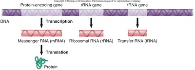
Messenger RNA (mRNA): Carries genetic information from DNA to ribosomes.
Ribosomal RNA (rRNA): Forms the core of ribosome structure and catalyzes protein synthesis.
Transfer RNA (tRNA): Brings amino acids to the ribosome during translation.
Protein Structure
Amino Acids and Peptide Bonds
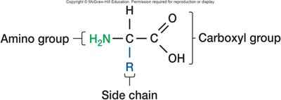
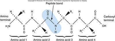
Proteins are polymers of amino acids linked by peptide bonds. Each amino acid contains a central carbon, an amino group (N-terminal), a carboxyl group (C-terminal), and a variable side chain (R group).
Side Chains: Determine the properties of amino acids (polar, non-polar, charged).
Peptide Bonds: Form between the carboxyl group of one amino acid and the amino group of another.
DNA Replication
Mechanism of DNA Replication
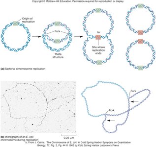
DNA replication is the process by which a cell duplicates its DNA before cell division. In most bacteria, DNA is circular and replication is bidirectional from a single origin.
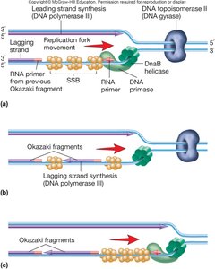
Replication Fork: The site where DNA is unwound and synthesis occurs.
Semiconservative Replication: Each new DNA molecule contains one original and one new strand.
Replication Machinery
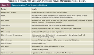
Multiple proteins and enzymes coordinate DNA replication. The table below summarizes key components in E. coli replication machinery.
Protein | Function |
|---|---|
DnaA (initiator protein) | Initiation of replication; binds origin of replication (oriC) |
DnaB | Helicase; separates DNA strands |
DNA gyrase | Relieves supercoiling ahead of replication fork |
SSB proteins | Bind single-stranded DNA after separation |
DnaC | Helicase loader; helps DnaB bind DNA |
DNA primase | Synthesizes RNA primer |
DNA polymerase III holoenzyme | Main DNA synthesis enzyme |
DNA polymerase I | Removes RNA primers; fills gaps with DNA |
Ribonuclease H | Removes RNA primers |
DNA ligase | Seals nicks; joins DNA fragments |
Topoisomerase IV | Separates chromosomes after replication |
Proofreading
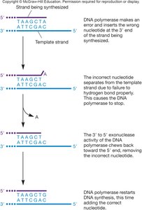
DNA polymerase III has exonuclease activity that removes mismatched bases from the 3' end of the growing strand, ensuring high fidelity during DNA replication.
Exonuclease Activity: Removes incorrect nucleotides.
Restart Synthesis: DNA polymerase resumes synthesis after correction.
Transcription
Transcription Process
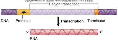
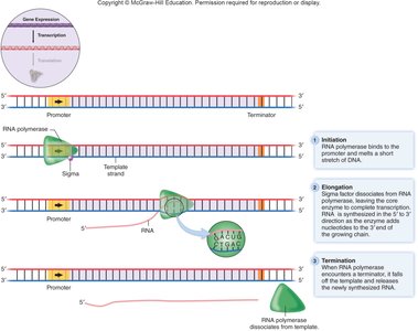
Transcription is the synthesis of RNA from a DNA template. RNA polymerase binds to the promoter region, synthesizes RNA in the 5' to 3' direction, and stops at the terminator.
Promoter: DNA sequence where RNA polymerase binds.
Terminator: DNA sequence signaling the end of transcription.
Direction: RNA is synthesized 5' to 3'.
Gene Expression in Bacteria
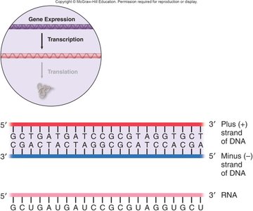
In bacteria, the RNA sequence is complementary and antiparallel to the DNA template strand. mRNA transcripts can be monocistronic (one gene) or polycistronic (multiple genes).
Minus (–) Strand: DNA template for RNA synthesis.
Plus (+) Strand: Complementary DNA strand; RNA sequence matches this strand except uracil replaces thymine.
Translation
Genetic Code and Translation
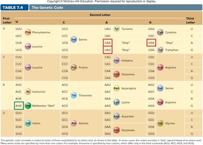
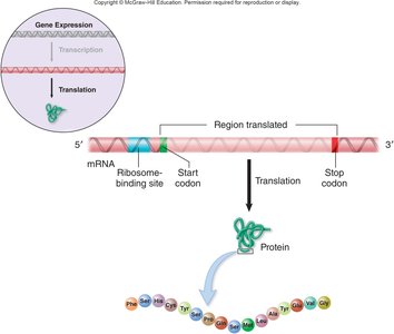
Translation is the process by which ribosomes interpret the information in mRNA to synthesize proteins. The genetic code consists of codons—three-nucleotide sequences that specify amino acids.
Codon: Three nucleotides in mRNA specify an amino acid.
Degeneracy: Multiple codons can code for the same amino acid.
Start Codon: AUG (methionine) initiates translation.
Stop Codons: UAA, UAG, UGA signal termination.
Codon | Amino Acid |
|---|---|
AUG | Methionine (Start) |
UUU | Phenylalanine |
UAA, UAG, UGA | Stop |
GCU | Alanine |
UUC | Phenylalanine |
CUU | Leucine |
Translation Process
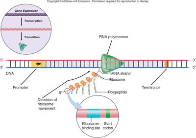
Translation involves initiation, elongation, and termination steps. Ribosomes move along mRNA, assembling amino acids into a polypeptide chain.
Initiation: Ribosome assembles at the start codon; tRNA brings the first amino acid.
Elongation: Ribosome moves along mRNA, adding amino acids via peptide bonds.
Termination: Ribosome reaches a stop codon; polypeptide is released.
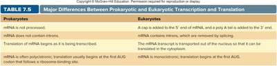
Differences Between Prokaryotic and Eukaryotic Gene Expression
Transcription and Translation Differences
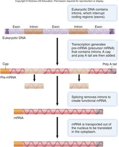
Eukaryotic transcription and translation differ from prokaryotic processes in several key ways, including mRNA processing, splicing, and ribosome structure.
Pre-mRNA: Eukaryotic mRNA is synthesized as a precursor and processed.
5' Cap: Methylated guanine added to stabilize and enhance translation.
Poly A Tail: ~200 adenine nucleotides added to the 3' end.
Splicing: Introns removed; exons joined.
Ribosomes: Eukaryotic ribosomes are 80S (40S and 60S subunits); prokaryotic ribosomes are 70S.
Monocistronic vs. Polycistronic: Eukaryotic mRNA is typically monocistronic; prokaryotic mRNA can be polycistronic.
Prokaryotes | Eukaryotes |
|---|---|
mRNA is not processed | 5' cap and poly A tail added |
mRNA does not contain introns | Introns removed by splicing |
Translation begins as mRNA is transcribed | mRNA transported out of nucleus for translation |
mRNA often polycistronic | mRNA typically monocistronic |
Gene Regulation
Types of Enzyme Regulation
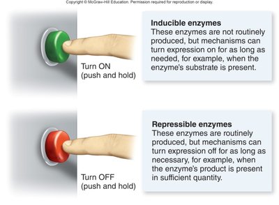
Cells regulate gene expression to conserve resources and respond to environmental changes. Enzymes can be constitutive, inducible, or repressible.
Constitutive Enzymes: Synthesized constantly; essential for central metabolism.
Inducible Enzymes: Produced only when needed (e.g., β-galactosidase when lactose is present).
Repressible Enzymes: Produced routinely; turned off when not required (e.g., amino acid synthesis).
Bacterial Gene Regulation Mechanisms
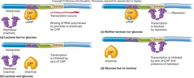
Bacteria use DNA-binding proteins to regulate transcription. Repressors and activators control gene expression by binding to operator regions.
Repressor: Blocks transcription (negative regulation).
Activator: Enhances transcription (positive regulation).
Induction: Inducer binds repressor, preventing DNA binding.
Repression: Corepressor enables repressor to bind DNA and block transcription.
Catabolite Repression and cAMP Regulation
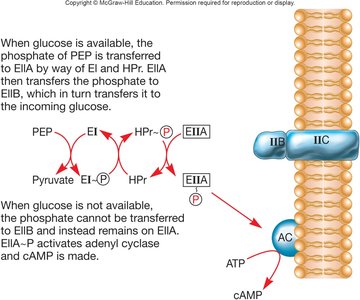
Catabolite repression allows preferential use of one carbon source over another. The phosphotransferase system regulates cAMP levels, which in turn affect gene expression.
Diauxic Growth: Biphasic growth pattern; lag occurs after preferred substrate is exhausted.
cAMP: Levels increase when glucose is not available, activating transcription of alternative pathways.
Eukaryotic Gene Regulation
Eukaryotic gene regulation is more complex, involving modification of chromosome structure, regulation of transcription initiation, pre-mRNA processing, and RNA interference (RNAi).
Chromosome Structure: Modification affects gene accessibility.
Transcription Initiation: Controlled by multiple regulatory elements.
RNAi: Small RNAs can silence gene expression post-transcriptionally.
