 Back
BackChapter 1 – Introduction to Microbiology: Comprehensive Study Notes
Study Guide - Smart Notes
 Tailored notes based on your materials, expanded with key definitions, examples, and context.
Introduction to Microbiology
What is Microbiology?
Microbiology is the scientific study of microorganisms, or microbes, which are often invisible to the naked eye. The term microbe includes both cellular, living microorganisms (such as bacteria, archaea, fungi, protists, and helminths) and nonliving/noncellular entities (such as viruses and prions). Microbes inhabit nearly every region of the planet, from deep-sea trenches to glaciers, and constitute at least half of Earth's life.
Prokaryotic cells: No nucleus, no membrane-bound organelles, typically unicellular, 10–100x smaller than eukaryotic cells. Examples: Bacteria and Archaea.
Eukaryotic cells: Possess a nucleus and membrane-bound organelles, can be unicellular or multicellular, 10–100x larger than prokaryotic cells. Examples: Fungi, Protists, Animals, Plants.
Microbes and Disease
Pathogens are microbes that cause disease. Of the thousands of known microbes, less than 1% are pathogenic to humans. Some pathogens always cause disease, while opportunistic pathogens cause disease only in weakened hosts.
Golden Age of Microbiology
Spontaneous Generation vs. Biogenesis
Early scientists debated whether life arose spontaneously from nonliving matter (spontaneous generation) or from existing life (biogenesis). Key experiments by Francesco Redi and Louis Pasteur disproved spontaneous generation, showing that life comes from existing life.
Francesco Redi: Demonstrated that maggots only appeared on meat when flies could access it.
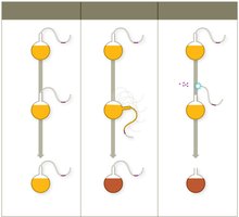
Louis Pasteur: Used S-necked flasks to show that microbes in the air, not spontaneous generation, caused contamination.
Germ Theory of Disease
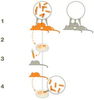
The germ theory of disease states that microbes cause infectious diseases. Robert Koch developed Koch's Postulates, a stepwise method to identify the causative agent of a disease:
The same organism must be present in every case of the disease, but not in healthy individuals.
The organism must be isolated and grown as a pure culture.
The isolated organism should cause the disease when inoculated into a susceptible host.
The organism must be re-isolated from the inoculated, diseased host.
Hand Hygiene and Aseptic Technique
Advances in aseptic technique, pioneered by Ignaz Semmelweis, Joseph Lister, and Florence Nightingale, greatly reduced infection rates in hospitals. Nosocomial infections (health-acquired infections) are prevented by practices such as hand washing, wearing gloves, sterilizing instruments, and decontaminating surfaces.
The Scientific Method
Steps in the Scientific Method
The scientific method is a systematic approach to investigation:
Observation
Hypothesis (tentative explanation)
Prediction
Experiment
Analysis
Conclusion (supports or rejects hypothesis)
Law: Precise statement or mathematical formula predicting a specific occurrence.
Theory: Hypothesis proven through repeated studies with consistent results; explains how and why something occurs.
Taxonomy and Classification
Taxonomic Hierarchy
Taxonomy is the study of how organisms are grouped by shared features. Early classification relied on morphology and physiology. Carl Linnaeus established the binomial nomenclature system and taxonomic hierarchy:
Domain
Kingdom
Phylum
Class
Order
Family
Genus
Species
Three domains: Bacteria, Archaea, Eukarya. Six kingdoms: Bacteria, Archaea, Plant, Protista, Animalia, Fungi.
Microbes: Friends or Foes
Microbial Diversity and Human Health
Microbes are abundant and diverse, with millions of species suspected. Most are harmless or beneficial, with only a minority being pathogenic. Microbes play essential roles in ecosystems and human health.
Host–Microbe Interactions
Symbiotic Relationships
Microbes and humans have evolved various symbiotic relationships:
Parasitism: Microbe harms the host (pathogens).
Mutualism: Both benefit.
Commensalism: No perceived benefit or harm to the host.
Normal Microbiota and the Human Microbiome Project
Normal Microbiota
The Human Microbiome Project aims to characterize all microbes in and on the human body. Normal microbiota includes bacteria, archaea, and eukaryotic microbes living in various body sites. Functions include training the immune system, producing vitamins, aiding digestion, and influencing mood and brain function.
Microbial antagonism: Normal microbiota protects by "crowding out" pathogens.
Some normal microbiota may include potential pathogens (e.g., Staphylococcus aureus).
Establishing Normal Microbiota
Babies are colonized by microbes during delivery and early interactions. Factors influencing microbiota development include delivery method and feeding type. Microbiota evolves from infancy to adulthood.
Host-Microbe Interactions and Human Evolution
Co-evolution and Disease Resistance
Humans and microbes have co-evolved. For example, carriers of the sickle cell gene have increased resistance to malaria, providing a survival advantage in regions where malaria is common.
Biofilms
Formation and Impact of Biofilms
Biofilms are sticky communities of microbes adhering to surfaces via polysaccharides. Biofilms develop in layers, providing protection and resistance to antibiotics and immune responses. They can form on teeth, contact lenses, medical devices, and more. The NIH estimates that 60–80% of human infectious diseases are due to biofilm-forming microbes.
Environmental and Industrial Uses for Microbes
Bioremediation
Microbes are harnessed for bioremediation to clean up toxic waste, such as degrading petroleum into CO2.
Culturing Microbes
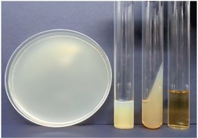
Growth Media
To study microbes, they are grown in laboratory environments using growth media (mixtures of nutrients). Agar is often used as a solidifying agent. Media types include broths, plates, slants, and deeps.
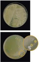
Aseptic Culture Technique
Aseptic techniques are used to prevent contamination when culturing microbes. This includes using sterile media, instruments, decontaminating surfaces, and protective clothing. The streak plate technique helps isolate colonies for study. A colony is a group of cells from a single parent cell; mixed cultures contain multiple colony types.

Staining Techniques
Stains and Dyes
Stains increase contrast for microscopic viewing. Basic dyes (positively charged) stain cells, while acidic dyes (negatively charged) stain backgrounds. Mordants fix dyes to specimens. Staining techniques include simple, structural, and differential stains.
Simple stains: Use one dye to determine size, shape, and arrangement.
Structural stains: Highlight specific structures (flagella, capsule, endospore).
Differential stains: Distinguish between cell types (e.g., Gram stain, acid-fast stain).
Gram Stain
The Gram stain classifies bacteria as Gram-positive (purple) or Gram-negative (pink) based on cell wall composition:
Gram-positive: Thick peptidoglycan, no outer membrane.
Gram-negative: Thin peptidoglycan, outer membrane rich in lipids.
Gram stain steps:
Crystal violet (primary stain)
Iodine (mordant)
Acetone-alcohol (decolorizer)
Safranin (counterstain)
Acid-Fast Staining
Acid-fast stain distinguishes cells with waxy cell walls (rich in mycolic acid). Acid-fast bacteria retain red dye after acid wash; important for detecting Mycobacterium and Nocardia.
Microscopy
Light Microscopy
Light microscopes use visible light and lenses to magnify specimens. The compound light microscope is most common, with objective and ocular lenses. Resolution is the ability to distinguish two points as separate; most compound microscopes achieve up to 1500x magnification and 200 nm resolution.
Oil Immersion
Oil immersion increases resolution at high magnification by reducing light scattering.
Types of Light Microscopy
Bright Field
Dark Field
Phase Contrast
Differential Interference Contrast
Electron Microscopy
Electron microscopes use electron beams for high-resolution imaging. Two main types:
Transmission Electron Microscope (TEM): Generates 2D images of internal structures.
Scanning Electron Microscope (SEM): Generates 3D images of surfaces.
Fluorescence in Microscopy
Fluorescence occurs when substances absorb UV light and emit visible light. Fluorochromes are dyes used to stain samples for fluorescence microscopy. Immunofluorescence uses fluorescent dyes linked to antibodies for specific identification of microbes.
Summary Table: Prokaryotic vs. Eukaryotic Cells
Feature | Prokaryotic Cells | Eukaryotic Cells |
|---|---|---|
Nucleus | No | Yes |
Membrane-bound Organelles | No | Yes |
Size | Smaller (0.1–5 µm) | Larger (10–100 µm) |
Examples | Bacteria, Archaea | Fungi, Protists, Animals, Plants |
Summary Table: Gram Stain Results
Cell Wall Type | Gram Stain Color | Peptidoglycan Layer | Outer Membrane |
|---|---|---|---|
Gram-positive | Purple | Thick | No |
Gram-negative | Pink | Thin | Yes |
Summary Table: Types of Growth Media
Type | Description |
|---|---|
Broth | Liquid medium for growing microbes |
Plate | Solid medium (agar) for isolating colonies |
Slant | Solid medium in a slanted tube for storage |
Deep | Solid medium in a deep tube for anaerobic growth |
