 Back
BackChapter 1: Introduction to Microbiology – Structured Study Notes
Study Guide - Smart Notes
 Tailored notes based on your materials, expanded with key definitions, examples, and context.
Introduction to Microbiology
Definition and Scope
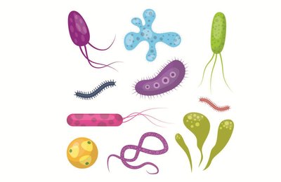
Microbiology is the scientific study of microorganisms, or microbes, which are often invisible to the naked eye. The field encompasses both cellular, living microorganisms (such as bacteria, archaea, fungi, protists, and helminths) and nonliving/noncellular entities (such as viruses and prions). Microbes inhabit nearly every region of the planet, from deep-sea trenches to glaciers, and constitute at least half of Earth's life.
Microbe: Any organism or particle studied in microbiology, including bacteria, archaea, fungi, protists, helminths, viruses, and prions.
Microbiology spans healthcare, agriculture, industry, and environmental sciences.
Humans rely on microbes for food production, medication synthesis, and environmental remediation.
Types of Microbes
Microbes are classified as either cellular (living) or noncellular (nonliving). Their cell type and characteristics are summarized below:
Microbe | Cell Type | Notes |
|---|---|---|
Bacteria | Prokaryotic | Unicellular; pathogenic and nonpathogenic |
Archaea | Prokaryotic | Unicellular; nonpathogenic; extremophiles |
Protists | Eukaryotic | Unicellular/multicellular; pathogenic and nonpathogenic |
Fungi | Eukaryotic | Unicellular/multicellular; pathogenic and nonpathogenic |
Helminths | Eukaryotic | Multicellular; parasitic worms |
Viruses | Noncellular | Infect animal, plant, or bacterial cells; DNA or RNA genome |
Prions | Noncellular | Infectious proteins; transmitted by transplant or ingestion |
Prokaryotic vs. Eukaryotic Cells
Microbes are divided into prokaryotic and eukaryotic cells. Prokaryotes (bacteria and archaea) evolved about 3.5 billion years ago and are unicellular. Eukaryotes include all multicellular organisms and some unicellular microorganisms (e.g., amoebae, yeast). The endosymbiotic theory explains the origin of eukaryotic cells.
Microbes and Disease
Pathogens and Opportunistic Pathogens
Pathogens are microbes that cause disease. Less than 1% of all microbes are pathogenic. Some pathogens always cause disease, while opportunistic pathogens cause disease only in weakened hosts.
History and Foundations of Microbiology
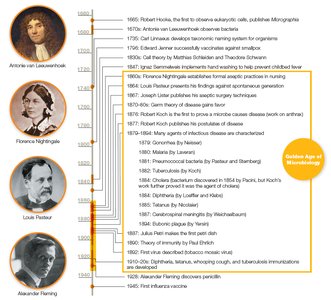
The Golden Age of Microbiology
The period from 1850 to 1920 saw major advances in microbiology, including innovations in microscopes, observations, and techniques to isolate and grow microbes.
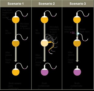
Spontaneous Generation vs. Biogenesis
Early scientists debated whether life arose from nonliving matter (spontaneous generation) or from existing life (biogenesis). Key experiments by Francesco Redi and Louis Pasteur disproved spontaneous generation, supporting biogenesis.
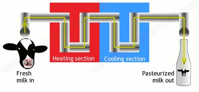
Pasteurization: Heating liquids to 50–60°C to kill microbes and prevent spoilage.
Pasteur developed vaccines against anthrax and rabies.
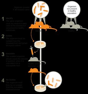
Germ Theory of Disease
The germ theory of disease states that microbes cause infectious diseases. Robert Koch developed techniques to identify the specific etiological agent of a disease, including staining and cultivation methods.
Aseptic Techniques and Hand Hygiene
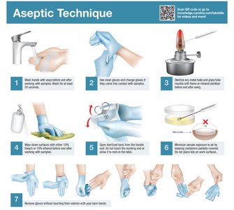
Development of Aseptic Techniques
Medical professionals such as Ignaz Semmelweis, Joseph Lister, and Florence Nightingale emphasized the importance of aseptic techniques to prevent healthcare-acquired infections (HAIs).
Hand washing
Wearing gloves
Sterilizing instruments
Decontaminating surfaces
The Scientific Method in Microbiology
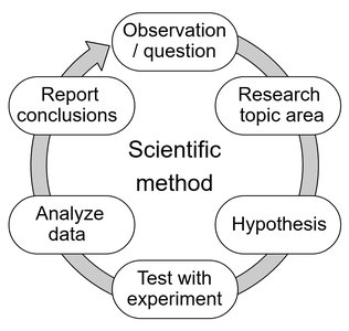
Principles and Steps
The scientific method guides investigations in microbiology. It involves forming a question, proposing a hypothesis, collecting and analyzing data, and drawing conclusions.
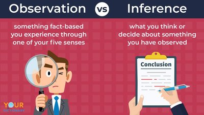
Observation: Data collected using senses or instruments.
Conclusion: Interpretation of observations.
Law vs. Theory
A scientific law is a precise statement or mathematical formula predicting a specific occurrence. A scientific theory is a hypothesis proven through many studies with consistent supporting conclusions. Laws predict what happens; theories explain how and why.
Taxonomy and Classification
Taxonomic Hierarchy
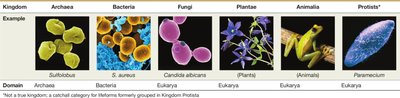
Taxonomy is the study of how organisms are grouped by shared features. The hierarchy includes eight ranks: Domain, Kingdom, Phylum, Class, Order, Family, Genus, Species.
Three domains: Bacteria, Archaea, Eukarya
Kingdoms: Animalia, Plantae, Fungi, Protista, Monera (older); Archaea and Bacteria (newer)
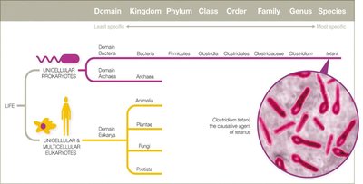
Scientific Names and Strains
Bacterial classification uses binomial nomenclature (Genus species, italicized). Strains are genetic variants of the same species, often denoted by numbers/letters (e.g., E. coli K-12).
Host–Microbe Interactions
Symbiotic Relationships
Microbes and humans have evolved various symbiotic relationships:
Parasitism: Microbe harms the host
Mutualism: Both benefit
Commensalism: No perceived benefit or cost
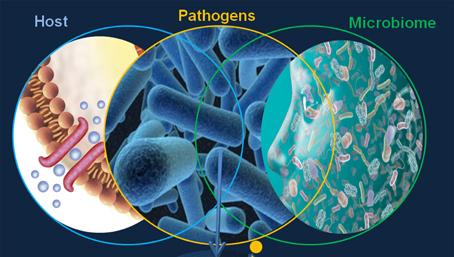
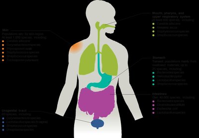
Normal Microbiota and Human Microbiome
The normal microbiota (normal flora) includes all microbes residing in and on the human body. The Human Microbiome Project aims to characterize these microbes. Functions include training the immune system, producing vitamins, aiding digestion, and possibly influencing mood and brain function.
Microbiota Disruptions and Transient Microbiota
Disruptions in normal microbiota, such as through antibiotic therapy, can increase infection risk by allowing opportunistic pathogens to establish infections. Transient microbiota are temporary and can be removed by hygiene.
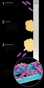
Biofilms
Formation and Significance
Biofilms are sticky communities of microbes that adhere to surfaces and form protective matrices. They are highly resistant to antibiotics and immune responses, and are implicated in 60–80% of human infectious diseases.
Environmental and Industrial Uses for Microbes
Bioremediation and Food Production
Microbes are used in bioremediation to clean up toxic waste and in food production (e.g., vinegar, beer, cheese, yogurt). They are also essential in drug production (e.g., penicillin, streptomycin).
Microbial Culture and Growth Media
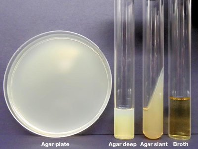
Growth Media Types
Growth media are nutrient mixtures supporting microbial growth in artificial settings. Types include broths, plates, slants, and deeps. Agar is a common solidifying agent.
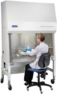
Aseptic Culture Techniques
Aseptic techniques are used to prevent contamination during microbial culture. Pure cultures are isolated using sterile media, instruments, and protective clothing. Biological safety cabinets minimize contamination risk.
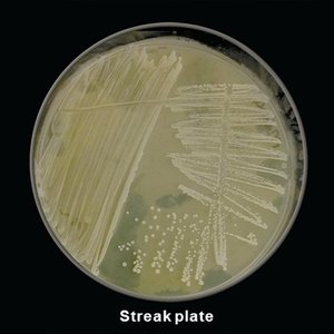
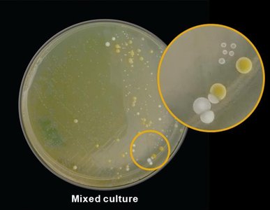
Streak Plate and Colony Isolation
The streak plate technique isolates colonies for study. Colonies are groups of cells from a single parent cell; mixed cultures contain multiple colony types.
Microscopy and Staining Techniques
Staining Methods
Stains increase contrast for microscopy. Basic dyes (positively charged) stain cells, while acidic dyes (negatively charged) stain backgrounds. Mordants fix dyes to specimens.
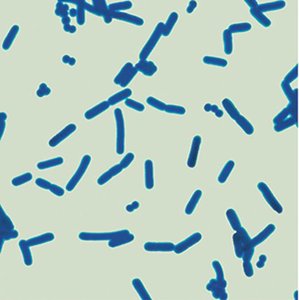
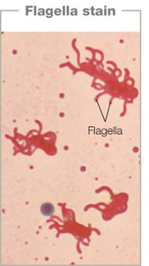
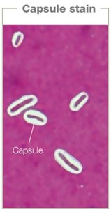
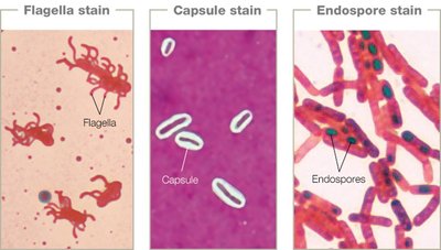
Simple, Structural, and Differential Stains
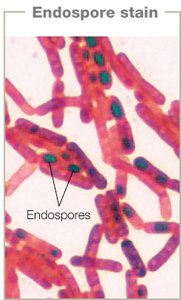
Simple stains use one dye to determine cell size, shape, and arrangement. Structural stains highlight features like flagella, capsules, and endospores. Differential stains (e.g., Gram, acid-fast) distinguish cell wall differences.
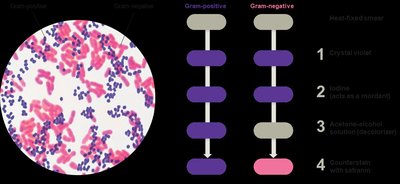
Gram Stain
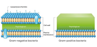
The Gram stain classifies bacteria as Gram-positive (thick peptidoglycan, no outer membrane) or Gram-negative (thin peptidoglycan, outer membrane rich in lipids). Gram-positive cells appear purple; Gram-negative cells appear pink.
Acid-Fast Staining
Acid-fast stains distinguish cells with waxy cell walls (rich in mycolic acid). Acid-fast bacteria retain red dye after acid wash; non–acid-fast cells do not. Used for Mycobacterium and Nocardia species.
Microscopy Types
Light Microscopy
Light microscopes use visible light and lenses to magnify specimens. Types include bright field, dark field, phase contrast, and differential interference contrast.
Microscopy Technique | Notes |
|---|---|
Bright Field | Sample appears dark on a bright background; requires staining. |
Dark Field | Sample appears light on a dark background; visualizes unstained specimens. |
Phase Contrast | Enhances contrast; visualizes live or dead specimens. |
DIC (Nomarski) | Provides false 3D appearance using polarized light. |
Electron Microscopy
Electron microscopes use electron beams for high-resolution imaging. Transmission electron microscopes (TEM) provide 2D images of internal structures; scanning electron microscopes (SEM) provide 3D images of surfaces.
Fluorescence Microscopy
Fluorescence occurs when substances absorb UV light and emit visible light. Fluorochromes stain samples for easy detection. Immunofluorescence uses antibodies linked to fluorescent dyes for specific identification.
Clinical Case Example: Cholera
Summary and Key Points
Cholera is an acute diarrheal illness caused by Vibrio cholerae, a waterborne bacterium. Proper sewage management reduces cases by limiting contamination. V. cholerae colonizes copepods in water, forming symbiotic relationships. Rugose colonies are more resistant to harsh conditions than smooth colonies.
*Additional info: The notes have been expanded with academic context and examples for clarity and completeness.*
