 Back
BackChapter 1
Study Guide - Smart Notes
 Tailored notes based on your materials, expanded with key definitions, examples, and context.
Chapter 1: The Microbial World
1.1 Microorganisms: Tiny Titans of the Earth
Microorganisms, or microbes, are life forms too small to be seen by the naked eye. They are highly diverse in form and function, inhabiting every environment that supports life. Most are single-celled, but some form complex structures or multicellular arrangements. Microbes often live in communities, interacting with each other and their environment.
Definition: Microorganisms are microscopic life forms, including bacteria, archaea, fungi, protists, and viruses.
Diversity: Microbes vary in morphology, metabolism, and ecological roles.
Habitats: Found in soil, water, air, and extreme environments.
Community: Microbial communities are groups of interacting microbes.
Domains: Three distinct lineages: Bacteria (prokaryotic), Archaea (prokaryotic), Eukarya (eukaryotic).
LUCA: All domains descended from the Last Universal Common Ancestor.
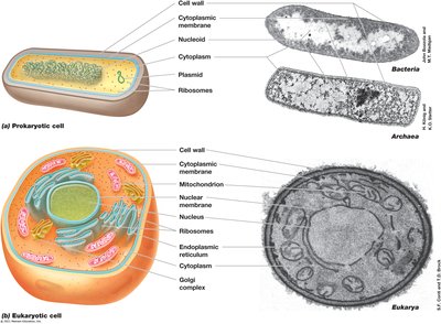
1.2 Structure and Activities of Microbial Cells
The cell is the fundamental unit of life, acting as a compartment that interacts with its environment. All cells share basic structural features, but differences exist between prokaryotic and eukaryotic cells.
Common Structures: Cytoplasmic membrane, cytoplasm, ribosomes, cell wall (in some microbes).
Prokaryotes: Bacteria and Archaea; lack membrane-bound organelles and nucleus.
Eukaryotes: Plants, animals, algae, protozoa, fungi; contain organelles and nucleus.
Genome: Full set of genes; prokaryotes usually have a single circular chromosome, eukaryotes have linear chromosomes.
Plasmids: Extra-chromosomal DNA in prokaryotes, often conferring special properties.
Cell Activities: Metabolism, growth, DNA replication, transcription, translation, motility, differentiation, communication, evolution.
1.3 Cell Size and Morphology
Microbial cells exhibit a wide range of sizes and shapes. Cell morphology affects nutrient uptake, growth rates, and evolutionary adaptations.
Size: Prokaryotes range from 0.2 μm to 600+ μm; most are 0.5–10 μm. Eukaryotes are typically 5–100 μm.
Surface-to-Volume Ratio: Smaller cells have higher ratios, supporting efficient nutrient exchange and faster growth.
Major Morphologies: Coccus (spherical), rod/bacillus (cylindrical), spirillum (flexible spiral), spirochete (rigid spiral), appendaged, irregular/asymmetrical, clusters.
Organism | Characteristics | Morphology | Size (μm) | Cell Volume (μm³) | Volume Compared to E. coli |
|---|---|---|---|---|---|
Thiomargarita namibiensis | Sulfur chemolithotroph | Cocci in chains | 750 | 200,000,000 | 100,000,000× |
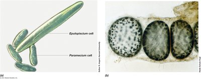
Epulopiscium fishelsoni | Chemoorganotroph | Rods with tapered ends | 80 × 600 | 3,000,000 | 1,500,000× |
Escherichia coli | Chemoorganotroph | Rods | 1 × 2 | 2 | 1× |
Mycoplasma pneumoniae | Pathogenic bacterium | Pleomorphic | 0.2 | 0.005 | 0.0025× |
Ultra-small bacteria | Uncultured, variable | Variable | <0.2 | 0.009 | 0.0045× |
1.4 An Introduction to Microbial Life
Microbial life is classified into three domains: Bacteria, Archaea, and Eukarya. Viruses, though not cellular, are important biological entities.
Bacteria: Prokaryotes, usually undifferentiated single cells, 80+ phylogenetic lineages.
Archaea: Prokaryotes, five well-described phyla, often extremophiles, no known pathogens.
Eukarya: Includes plants, animals, fungi, protists; first were unicellular, at least six kingdoms.
Viruses: Obligate parasites, not cells, replicate only within host cells, classified by structure and genome.
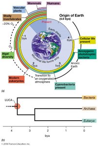
1.5 Microorganisms and the Biosphere
Microorganisms have shaped Earth's biosphere since its origin. They are the oldest forms of life and constitute a major fraction of Earth's biomass. Extremophiles thrive in habitats too harsh for other life forms.
History: Earth is 4.6 billion years old; first cells appeared 3.8–4.3 billion years ago.
Anoxic Atmosphere: Early Earth lacked O2; only anaerobic metabolisms.
Phototrophs: First anoxygenic phototrophs ~3.6 bya; cyanobacteria (oxygenic) ~2.6 bya.
Extremophiles: Microbes living in extreme conditions (hot springs, glaciers, high salt, acidity, pressure).
Microbial Ecology: Study of how microbes affect plants, animals, and ecosystems.
Extreme | Descriptive Term | Genus, Species | Domain | Habitat | Minimum | Optimum | Maximum |
|---|---|---|---|---|---|---|---|
Temperature High | Hyperthermophile | Methanopyrus kandleri | Archaea | Hydrothermal vents | 90°C | 106°C | 122°C |
Temperature Low | Psychrophile | Psychromonas ingrahamii | Bacteria | Sea ice | -12°C | 5°C | 10°C |
pH Low | Acidophile | Picrophilus oshimae | Archaea | Acidic hot springs | -0.06 | 0.7 | 4 |
pH High | Alkaliphile | Natronobacterium gregoryi | Archaea | Soda lakes | 8.5 | 10 | 12 |
Pressure | Barophile | Moritella yayanosii | Bacteria | Deep ocean sediments | 500 atm | 700 atm | >1000 atm |
Salt | Halophile | Halobacterium salinarum | Archaea | Salterns | 15% | 25% | 32% (saturation) |
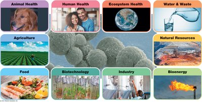
1.6 The Impact of Microorganisms on Human Society
Microorganisms play crucial roles in human health, agriculture, industry, and environmental processes. They can be both beneficial and harmful.
Disease Agents: Cause infectious diseases; control achieved through vaccination, antibiotics, water treatment, and food safety.
Agriculture: Nitrogen-fixing bacteria, cellulose-degrading microbes, gut microbiome, vitamin synthesis.
Food: Cause spoilage and foodborne disease; also used in fermentation (cheese, yogurt, sauerkraut, bread, alcohol).
Industry: Industrial microbiology (pharmaceuticals, brewing), biotechnology (genetically engineered microbes), biofuels, wastewater treatment, bioremediation, biofilms.
1.7 Microscopy and the Origins of Microbiology
Microbiology began with the invention of the microscope. Early pioneers like Robert Hooke and Antoni van Leeuwenhoek made foundational discoveries.
Robert Hooke: First to describe microbes (molds).
Antoni van Leeuwenhoek: First to see bacteria using a light microscope.
Microscopy: Magnification and resolution are key concepts; several types exist (bright-field, phase-contrast, dark-field, fluorescence).
Compound Light Microscope: Uses visible light, condenser, objective and ocular lenses; total magnification = objective × ocular.
Bright-field Scope: Visualizes specimens by contrast differences.
1.8 Improving Contrast in Light Microscopy
Staining techniques enhance contrast in microscopy, allowing better visualization of cellular structures.
Staining: Dyes bind to cellular materials; basic dyes are positively charged.
Simple Stain: Uses dried cells.
Differential Stains: Render different cells different colors; Gram stain distinguishes gram-positive (purple) and gram-negative (pink) bacteria.
Phase-Contrast Microscopy: Amplifies refractive index differences; dark cells on light background.
Dark-Field Microscopy: Light from sides; specimen appears light on dark background; good for motility.
Fluorescence Microscopy: Visualizes specimens that fluoresce; used in clinical diagnostics and microbial ecology.
1.9 Imaging Cells in Three Dimensions
Advanced microscopy techniques allow three-dimensional visualization of cells and their structures.
DIC Microscopy: Uses polarized light to create 3D appearance.
Confocal Scanning Laser Microscopy: Computerized fluorescent microscope with laser source; generates 3D images by focusing on single layers.
1.10 Probing Cell Structure: Electron Microscopy
Electron microscopes use electrons to image cells, providing much greater resolution than light microscopes.
Transmission Electron Microscopy (TEM): Visualizes structures at molecular level; specimen must be thin and stained.
Scanning Electron Microscopy (SEM): Scans specimen coated with heavy metal; only surface visualized; magnification up to 100,000×.
1.11 Pasteur and Spontaneous Generation
Louis Pasteur disproved the theory of spontaneous generation and made major contributions to microbiology.
Pasteur Flask Experiment: Showed life does not arise spontaneously from nonliving material.
Vaccines: Developed for anthrax, fowl cholera, rabies.
Fermentation: Demonstrated biological basis.
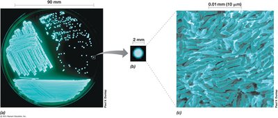
1.12 Koch, Infectious Disease, and Pure Cultures
Robert Koch established the link between microbes and infectious diseases, developed pure culture techniques, and formulated Koch’s postulates.
Koch’s Postulates: Criteria to definitively link cause and effect in infectious disease.
Solid Media: Enabled isolation of pure cultures.
Microbial Diversity: Observed differences in colonies and cell types.
1.13 Discovery of Microbial Diversity
Microbial diversity research focuses on nonmedical aspects and metabolic processes in soil and water.
Winogradsky: Linked specific bacteria to biogeochemical transformations; proposed chemolithotrophy.
Beijerinck: Developed enrichment culture technique; isolated aerobic nitrogen-fixing bacteria; first to observe a virus.
1.14 Molecular Basis of Life
Bacteria are excellent models for studying the fundamental nature of life, leading to advances in molecular biology, genetics, and biochemistry.
Universal Macromolecules: Certain molecules and reactions are universal across life.
DNA as Genetic Material: Experiments by Griffith, Avery-MacLeod-McCarty, Watson, Crick, Franklin established DNA as the basis of heredity.
1.15 Woese and the Tree of Life
Carl Woese used ribosomal RNA sequences to infer evolutionary relationships, leading to the three-domain classification of life.
Phylogenetic Tree: Depicts evolutionary history; root is LUCA.
Domains: Bacteria, Archaea, Eukarya.
Cultivation-Independent Methods: Reveal most microbes have not been cultured.
DNA Sequencing: Enables study of entire genomes and metagenomics.
Additional info: These notes expand on the provided lecture slides and textbook images, adding definitions, examples, and context for microbiology students. All included images are directly relevant to the adjacent content, reinforcing key concepts.
