 Back
Chapter 1: The Microbial World – Foundations and Applications
Study Guide - Smart Notes
 Tailored notes based on your materials, expanded with key definitions, examples, and context.
The Microbial World
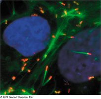
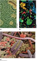
Microbial Communities
Microorganisms exist in complex communities, interacting with each other and their environment. These communities can be found in diverse habitats, including soil, water, and within host organisms. The study of microbial communities is essential for understanding ecosystem function, health, and disease.
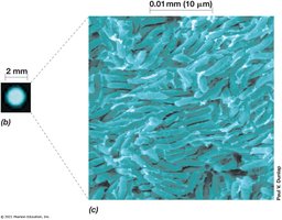
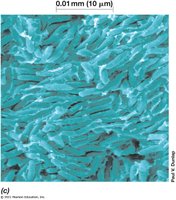
Microbial growth refers to the multiplication and replication of individual cells, resulting in an increase in cell numbers.
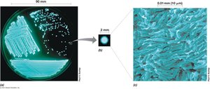
Each colony on a solid medium is typically derived from a single cell, known as a colony forming unit (CFU).
Ideally, all cells in a colony share the same genetic information.
Applications of Microbiology
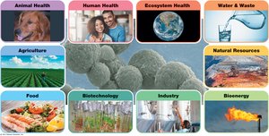
Microbiology has broad applications across multiple fields, impacting human health, agriculture, industry, and environmental management. Microbes are utilized in biotechnology, food production, wastewater treatment, and bioenergy.
Human and animal health: Microbes play roles in disease, immunity, and symbiosis.
Agriculture: Microbes contribute to soil fertility and plant growth.
Industry: Used in fermentation, bioremediation, and production of pharmaceuticals.
Bioenergy: Microbes convert waste into usable energy.
Microbial Cell Structure and Function
Microbial Cells and Colonies
Microbial cells are the basic units of life, capable of independent growth and reproduction. Colonies on solid media are visible clusters of cells derived from a single ancestor.
Replication: Microbial growth is measured by the increase in cell numbers.
Genetic uniformity: Cells in a colony are genetically identical unless mutations occur.
Cell Structure: Prokaryotes vs. Eukaryotes
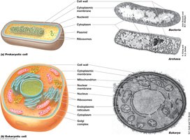
Microbial cells are classified as prokaryotic or eukaryotic based on their structural features. Prokaryotes lack a nucleus, while eukaryotes possess a membrane-bound nucleus and organelles.
Prokaryotes: Include Bacteria and Archaea; DNA is not enclosed in a nucleus and is typically not associated with histone proteins (except Archaea have histone-like proteins).
Eukaryotes: Include fungi, protozoa, algae; DNA is enclosed in a nucleus and associated with histones.
Plasmids: Extra-chromosomal DNA in bacteria, often carrying antibiotic resistance genes.
Cytoplasm: Site of metabolism, compartmentalized in eukaryotes.
Organelles: Mitochondria (energy, amino/lipid metabolism), Golgi apparatus (protein modification), Endoplasmic reticulum (protein and lipid synthesis).
Properties of Microbial Cells
Metabolism, Growth, and Evolution
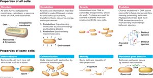
Microbial cells exhibit fundamental properties such as metabolism, growth, and evolution. Metabolism encompasses all chemical reactions within a cell, enabling energy production and biosynthesis.
Metabolism: Sum of all chemical reactions; includes catabolism (energy production) and anabolism (biosynthesis).
Growth: Cells replicate their DNA and divide, increasing population size.
Evolution: Genetic changes accumulate over time, leading to adaptation and speciation.
Anfibolic pathways: Pathways that serve both catabolic and anabolic functions, e.g., Krebs Cycle.
Cell Size and Volume
Microbial cells vary greatly in size and volume, influencing their metabolic rates and ecological roles. The size of a cell affects its surface area-to-volume ratio, which is critical for nutrient uptake and waste removal.
Largest bacteria: Thiomargarita namibiensis and Epulopiscium fishelsoni.
Smallest bacteria: Pelagibacter ubique and Mycoplasma pneumoniae.
Cell size is measured in micrometers (μm); volume in cubic micrometers (μm3).
Organism | Characteristics | Morphology | Size (μm) | Cell volume (μm3) | Volume compared to E. coli |
|---|---|---|---|---|---|
Thiomargarita namibiensis | Sulfur chemolithotroph | Coccus | 100–300 | 2,000,000 | 3,000–6,000 |
Epulopiscium fishelsoni | Chemoorganotroph | Rod with tapered ends | 80 × 600 | 1,000,000 | 1,500–3,000 |
Escherichia coli | Chemoorganotroph | Rod | 2 × 0.6 | 0.6 | 1 |
Pelagibacter ubique | Chemoorganotroph | Rod | 0.2 × 0.5 | 0.01 | 0.016 |
Mycoplasma pneumoniae | Chemoorganotroph, no cell wall | Variable | 0.2 | 0.004 | 0.0066 |
Additional info: Table includes more organisms and their characteristics. | |||||

Surface Area-to-Volume Relationships
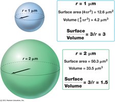
The surface area-to-volume ratio is a key factor in cell physiology. Smaller cells have a higher ratio, facilitating efficient nutrient uptake and waste removal.
Surface area of a sphere:
Volume of a sphere:
Surface area-to-volume ratio:
As cell size increases, the ratio decreases, limiting metabolic efficiency.
Cell Morphologies
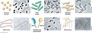
Microbial cells exhibit diverse morphologies, including cocci (spherical), rods (bacilli), spirilla (spiral), and filamentous forms. Morphology can influence motility, nutrient acquisition, and ecological niche.
Coccus: Spherical shape.
Rod: Cylindrical shape.
Spirillum: Spiral shape.
Filamentous: Long, thread-like cells.
Other forms: Spirochetes, stalked, hyphal, budding, and appendaged cells.
Microbial Diversity and Evolution
Microorganisms in the Environment
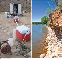
Microbes are found in every environment on Earth, including extreme habitats such as anoxic groundwater, hot springs, and saline lakes. Their adaptability is due to diverse metabolic pathways and genetic variation.
Sampling techniques are used to study microbial diversity in natural environments.
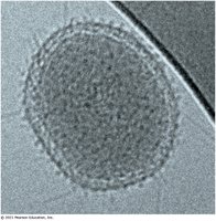
Microbes can pass through filters with very small pore sizes, indicating their minute size.
Extremophiles
Extremophiles are microorganisms that thrive in extreme conditions of temperature, pH, pressure, or salinity. They are classified based on their optimal growth conditions.
Extreme | Description | Genus/Species | Domain | Habitat | Minimum | Optimum | Maximum |
|---|---|---|---|---|---|---|---|
Temperature | Hyperthermophile | Archaeon Pyrolobus fumarii | Archaea | Hydrothermal vent | 90°C | 106°C | 113°C |
pH | Acidophile | Picrophilus oshimae | Archaea | Acidic hot springs | 0.06 | 0.7 | 4 |
Pressure | Barophile | Halomonas salaria | Bacteria | Saline | 0% | 20% | 32% |
Additional info: Table includes more extremophiles and their characteristics. | |||||||

Microbial Evolution and the Tree of Life
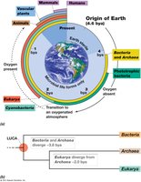
Microorganisms have played a central role in the evolution of life on Earth. The phylogenetic tree of life is based on ribosomal RNA (rRNA) sequences, revealing three domains: Bacteria, Archaea, and Eukarya.
LUCA: Last Universal Common Ancestor, from which all cellular life diverged.
Domains: Bacteria, Archaea, Eukarya.
Microbial evolution is traced through molecular and fossil evidence.
Historical Foundations of Microbiology
Early Microscopy and Discovery of Microbes
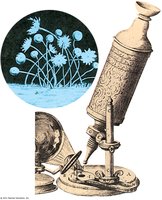
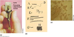
The development of microscopy was crucial for the discovery and study of microorganisms. Early pioneers such as Robert Hooke and Antonie van Leeuwenhoek made significant contributions to the field.
Robert Hooke: First to describe cells and microorganisms using a compound microscope.
Van Leeuwenhoek: Improved lens technology and observed single-celled organisms.
Defeat of Spontaneous Generation
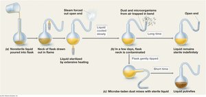
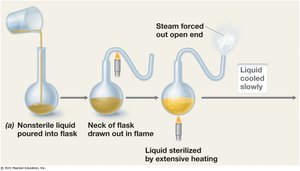
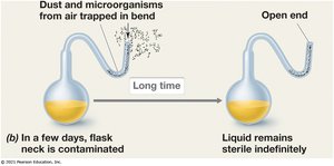
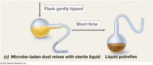
Louis Pasteur's experiments disproved the theory of spontaneous generation, demonstrating that life arises from pre-existing cells. His swan-necked flask experiment showed that sterilized broth remains free of microbes unless exposed to contaminated air.
Spontaneous generation: The false belief that life could arise from non-living matter.
Pasteur's experiment: Used flasks with long necks to prevent airborne microbes from contaminating sterile broth.
Koch's Postulates
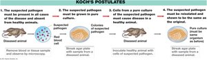
Robert Koch established a set of criteria, known as Koch's postulates, for proving the causal relationship between a microorganism and a disease. These postulates are fundamental to medical microbiology and infectious disease research.
1. The suspected pathogen must be present in all cases of the disease and absent from healthy animals.
2. The pathogen must be grown in pure culture.
3. Cells from the pure culture must cause disease in a healthy animal.
4. The pathogen must be reisolated and shown to be the same as the original.
Conclusion
Chapter 1 provides a comprehensive overview of the microbial world, including the structure and function of microbial cells, their diversity, evolution, and historical foundations. Understanding these concepts is essential for further study in microbiology and its applications in health, industry, and environmental science.
