 Back
BackChapter 12: Host Defenses I – Overview and Innate Defenses
Study Guide - Smart Notes
 Tailored notes based on your materials, expanded with key definitions, examples, and context.
Host Defenses: An Overview
Three Lines of Host Defense
The human body employs a multilevel network of defenses to protect against microbial invasion. These defenses are categorized into three lines: the first and second lines are innate (nonspecific), while the third line is adaptive (specific).
First Line of Defense: Physical, chemical, and microbiota barriers that prevent pathogen entry.
Second Line of Defense: Cellular and chemical responses such as phagocytosis, inflammation, fever, and antimicrobial products.
Third Line of Defense: Adaptive immunity involving lymphocytes (B and T cells) that provide specific, long-term protection.
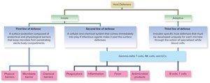
Example: The skin acts as a physical barrier (first line), while phagocytes and inflammation (second line) respond if pathogens breach the skin. If infection persists, adaptive immunity (third line) is activated.
Immune Recognition and Surveillance
Markers and Immune System Function
Immune cells distinguish self from nonself by recognizing molecular markers (antigens) on cell surfaces. This recognition is crucial for targeting pathogens while sparing host tissues.
Markers (Antigens): Molecules (proteins/sugars) on cell surfaces used for identification.
Pathogen-Associated Molecular Patterns (PAMPs): Common microbial markers recognized by the immune system.
Pattern Recognition Receptors (PRRs): Host cell receptors that detect PAMPs and initiate immune responses.
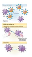
Example: White blood cells use PRRs to detect PAMPs on bacteria, leading to phagocytosis and destruction of the invader.
Body Systems Involved in Immunity
Lymphatic and Circulatory Systems
The immune system is a diffuse network involving the circulatory and lymphatic systems, which transport immune cells and facilitate communication between compartments.
Circulatory System: Screens body compartments via circulating white blood cells.
Lymphatic System: Network of vessels, nodes, and organs that transport lymph and house immune cells.
Connection: Lymphatic vessels drain into the circulatory system near the heart, allowing immune cells to circulate throughout the body.
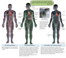
Example: Lymph nodes filter lymph and provide sites for immune cell activation.
Lymphatic System Structure and Function
Lymphatic Organs and Tissues
The lymphatic system includes primary and secondary organs essential for immune cell development and function.
Primary Organs: Red bone marrow (site of blood cell production and B cell maturation), thymus (site of T cell maturation).
Secondary Organs: Lymph nodes, spleen, and associated lymphoid tissues (sites of immune cell activation and response).
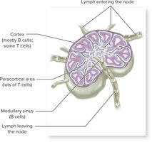
Example: Lymph nodes contain regions rich in B and T cells, facilitating immune surveillance and response to pathogens.
Blood Cells in Immunity
Hematopoiesis and Leukocyte Differentiation
Blood cells originate from pluripotent stem cells in the bone marrow and differentiate into various lineages, including those critical for immunity.
Granulocytes: Neutrophils, eosinophils, basophils (contain granules, involved in innate responses).
Agranulocytes: Monocytes (differentiate into macrophages and dendritic cells), lymphocytes (B and T cells, NK cells).
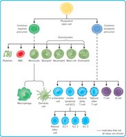
Example: Neutrophils are the most abundant phagocytes and are first responders to infection.
Mononuclear Phagocyte System (MPS)
Structure and Function
The MPS is a network of phagocytic cells (macrophages, dendritic cells) distributed throughout tissues, providing a first line of defense against pathogens.
Reticulum: Connective tissue network supporting immune cells.
Locations: Thymus, lymph nodes, spleen, tonsils, mucosal tissues.
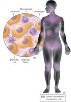
Example: Macrophages in the liver (Kupffer cells) and skin (Langerhans cells) are specialized tissue-resident phagocytes.
Histiocytes: Tissue-Resident Macrophages
Specialized Macrophages in Tissues
Histiocytes are differentiated macrophages and dendritic cells that reside permanently in specific tissues, providing localized immune defense.
Kupffer Cells: Liver macrophages.
Alveolar Macrophages: Lungs.
Langerhans Cells: Skin.
Microglia: Brain.
Example: Langerhans cells in the skin detect and present antigens to T cells, initiating immune responses.
First Line of Defense: Barriers
Physical and Chemical Barriers
The first line of defense consists of anatomical and physiological barriers that prevent pathogen entry.
Physical Barriers: Intact skin, mucous membranes, cilia, hair, and flushing actions (tears, saliva, urine).
Chemical Barriers: Lysozyme in tears/saliva, acidic pH of skin and stomach, antimicrobial peptides, sebum, and sweat.
Microbiota Barrier: Normal microbiota compete with pathogens for nutrients and space.
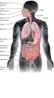
Example: Lysozyme in tears hydrolyzes bacterial cell walls, preventing eye infections.
Respiratory Tract Defenses
Ciliary Escalator and Mucus
The respiratory tract is protected by mucus and ciliated epithelial cells that trap and expel pathogens.
Nasal Hair: Traps large particles.
Ciliary Escalator: Moves trapped particles toward the pharynx for removal.
Sneeze/Cough Reflexes: Expel irritants and pathogens.
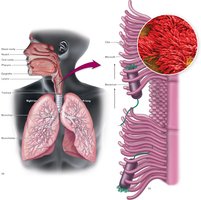
Example: Cilia in the trachea move mucus and trapped microbes upward, preventing lung infections.
Second Line of Defense: Cellular and Chemical Responses
Phagocytosis
Phagocytosis is a key process in innate immunity, involving the engulfment and destruction of pathogens by specialized cells.
Phagocytes: Neutrophils, monocytes/macrophages, dendritic cells.
Steps: Chemotaxis, adhesion, ingestion, phagosome formation, phagolysosome formation, destruction, excretion.
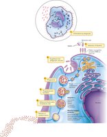
Example: Neutrophils migrate to infection sites, engulf bacteria, and destroy them using reactive oxygen species and enzymes.
Inflammatory Response
Stages and Functions of Inflammation
Inflammation is a complex, localized response to tissue injury or infection, characterized by redness, heat, swelling, pain, and loss of function.
Stages: Vasoconstriction, vasodilation, increased vascular permeability, leukocyte migration, tissue repair.
Functions: Attract immune cells, contain and eliminate pathogens, initiate tissue repair.
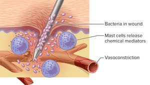
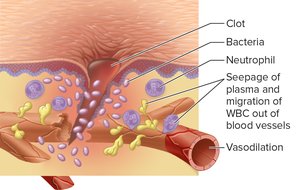
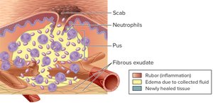
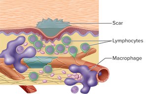
Example: Neutrophils and macrophages clear debris and pathogens, while fibroblasts repair tissue.
Diapedesis and Chemotaxis
Leukocyte Migration to Infection Sites
Diapedesis is the process by which white blood cells exit blood vessels and enter tissues, guided by chemotactic signals to sites of infection or injury.
Diapedesis: Movement of leukocytes through vessel walls.
Chemotaxis: Directed migration toward chemical signals released by pathogens or damaged tissues.
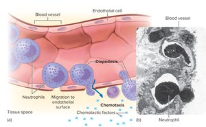
Example: Neutrophils follow chemotactic gradients to accumulate at infection sites.
Antimicrobial Products
Interferons
Interferons are small proteins produced by host cells in response to viral infection, with roles in immune regulation and antiviral defense.
Types: IFN-α, IFN-β (produced by lymphocytes, fibroblasts, macrophages); IFN-γ (produced by T cells).
Functions: Inhibit viral replication, activate immune cells, suppress tumor growth.
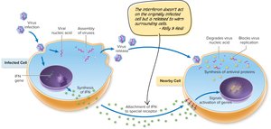
Example: Interferons induce antiviral proteins in neighboring cells, blocking viral replication.
Complement System
The complement system is a cascade of over 30 blood proteins that enhance immune responses and directly destroy pathogens.
Pathways: Classical (antibody-dependent), alternative (antibody-independent).
Stages: Initiation, activation/cascade, polymerization, membrane attack (formation of membrane attack complex, MAC).
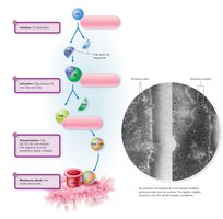
Example: The MAC forms pores in microbial membranes, leading to cell lysis.
Antimicrobial Peptides
Short proteins (e.g., defensins) that disrupt microbial membranes, leading to cell lysis. They are part of both innate and adaptive immunity and are being explored as therapeutic agents.
Mechanism: Insert into membranes, form pores, cause cell death.
Example: Human defensins are produced by epithelial cells and neutrophils to kill bacteria and fungi.
