 Back
BackChapter 3 Microbiology: Microscopy, Staining, and Microbial Size Study Guide
Study Guide - Smart Notes
 Tailored notes based on your materials, expanded with key definitions, examples, and context.
Q1. If a microbe measures 10 μm in length, how long is it in nanometers? In mm? Based on its size, what group do you suspect this agent belongs to? How would you go about confirming that?
Background
Topic: Microbial Size and Classification
This question tests your understanding of metric unit conversions (micrometers to nanometers and millimeters), and your ability to relate microbial size to classification (e.g., bacteria, protozoa, viruses). It also asks you to think about how to confirm the identity of a microbe based on its size.
Key Terms and Formulas
Micrometer (μm): meters
Nanometer (nm): meters
Millimeter (mm): meters
Conversion factors:

Step-by-Step Guidance
Start by converting 10 μm to nanometers using the conversion factor .
Next, convert 10 μm to millimeters using the conversion factor .
Compare the size (10 μm) to the typical size ranges of different microbial groups (e.g., bacteria, protozoa, fungi, viruses). Refer to a size chart if needed.
Think about what laboratory methods (such as microscopy, staining, or culturing) could be used to confirm the identity of the microbe based on its size and morphology.
Try solving on your own before revealing the answer!
Q2. What does it mean when a microscope has a resolution of 0.2 nm?
Background
Topic: Microscopy - Resolution
This question is about understanding the concept of resolution in microscopy, specifically what it means for a microscope to have a certain resolving power.
Key Terms and Formulas
Resolution: The ability of a microscope to distinguish two points as separate entities. The smaller the value, the better the resolution.
Resolving Power: The minimum distance between two points that can still be distinguished as separate.
Step-by-Step Guidance
Define what is meant by 'resolution' in the context of microscopy.
Interpret what a resolution of 0.2 nm means in practical terms (i.e., what size objects can be distinguished).
Consider what types of microscopes are capable of achieving this level of resolution.
Try solving on your own before revealing the answer!
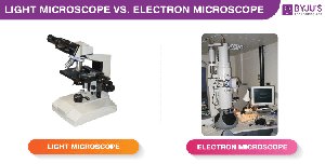
Q3. Why do electron microscopes have greater resolution than light microscopes?
Background
Topic: Microscopy - Electron vs. Light Microscopes
This question tests your understanding of the physical principles behind different types of microscopes and why electron microscopes can resolve smaller structures than light microscopes.
Key Terms and Formulas
Wavelength: The distance between two consecutive peaks of a wave. Shorter wavelengths allow for higher resolution.
Resolution formula: , where is the minimum resolvable distance, is the wavelength, and is the numerical aperture.
Step-by-Step Guidance
Recall that the resolution of a microscope is limited by the wavelength of the radiation used to view the specimen.
Compare the wavelengths of visible light (used in light microscopes) and electrons (used in electron microscopes).
Explain how the shorter wavelength of electrons allows electron microscopes to resolve much smaller structures than light microscopes.
Try solving on your own before revealing the answer!
Q4. Why doesn’t a negative stain color a cell?
Background
Topic: Staining Techniques in Microbiology
This question is about understanding the principles behind negative staining and why it results in a different appearance compared to positive stains.
Key Terms and Formulas
Negative stain: A staining technique in which the background is stained, leaving the cells colorless.
Cell surface charge: Most bacterial cell surfaces are negatively charged.
Step-by-Step Guidance
Recall the charge properties of bacterial cell surfaces and the dyes used in negative staining.
Explain how the interaction (or lack thereof) between the dye and the cell surface leads to the observed staining pattern.
Describe what you would expect to see under the microscope after performing a negative stain.
Try solving on your own before revealing the answer!
Q5. Why is the Gram stain so useful?
Background
Topic: Differential Staining - Gram Stain
This question tests your understanding of the Gram stain technique and its importance in microbiology for classifying bacteria.
Key Terms and Formulas
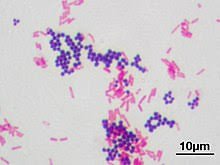
Gram-positive bacteria: Bacteria that retain the crystal violet stain and appear purple.
Gram-negative bacteria: Bacteria that do not retain the crystal violet stain and appear pink/red after counterstaining.
Cell wall structure: The main reason for the difference in staining is the structure of the bacterial cell wall.
Step-by-Step Guidance
Recall the steps of the Gram stain and the differences in cell wall structure between Gram-positive and Gram-negative bacteria.
Explain how these differences result in different staining outcomes.
Discuss why this distinction is important for diagnosis and treatment in clinical microbiology.
Try solving on your own before revealing the answer!
