 Back
Chapter 3 Microbiology Study Guidance: Microscopy, Staining, and Microbial Classification
Study Guide - Smart Notes
 Tailored notes based on your materials, expanded with key definitions, examples, and context.
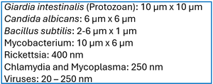
Q1. If a microbe measures 10 μm in length, how long is it in nanometers? In mm? Based on its size, what group do you suspect this agent belongs to? How would you go about confirming that?
Background
Topic: Microbial Size and Classification
This question tests your understanding of metric conversions (micrometers to nanometers and millimeters), and how microbial size relates to classification (e.g., bacteria, protozoa, viruses).
Key Terms and Formulas
Micrometer (m): meters
Nanometer (nm): meters
Millimeter (mm): meters
Conversion: ;
Step-by-Step Guidance
Start by converting 10 m to nanometers using the conversion factor: .
Next, convert 10 m to millimeters using the conversion factor: .
Compare the size to typical microbial groups (see the provided chart for reference).
Think about what group (bacteria, protozoa, fungi, virus) this size fits best.
Consider what laboratory methods (e.g., microscopy, staining) could confirm the identity of the microbe.

Try solving on your own before revealing the answer!
Final Answer:
10 m = 10,000 nm; 10 $\mu$m = 0.01 mm. Based on the size, this microbe is likely a protozoan or a large bacterium. To confirm, you would use microscopy and possibly staining techniques to observe its morphology and characteristics.
Q2. What does it mean when a microscope has a resolution of 0.2 nm?
Background
Topic: Microscopy and Resolution
This question tests your understanding of the concept of resolution in microscopy, which is the ability to distinguish two points as separate.
Key Terms and Formulas
Resolution: The minimum distance at which two points can be distinguished as separate.
Unit: Nanometers (nm)
Step-by-Step Guidance
Define what resolution means in the context of microscopy.
Interpret what a value of 0.2 nm indicates about the microscope's capabilities.
Consider what types of microscopes can achieve this level of resolution.
Think about what kinds of specimens can be visualized at this resolution.
Try solving on your own before revealing the answer!
Final Answer:
A resolution of 0.2 nm means the microscope can distinguish two points that are 0.2 nm apart. This is typical of electron microscopes, allowing visualization of very small structures such as viruses and molecular complexes.
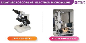
Q3. Why do electron microscopes have greater resolution than light microscopes?
Background
Topic: Microscopy Types and Resolution
This question tests your understanding of the physical principles behind different types of microscopes and why electron microscopes outperform light microscopes in resolution.
Key Terms and Formulas
Resolution (): , where is wavelength and is numerical aperture.
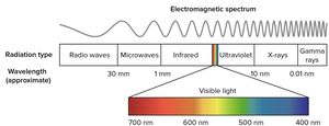
Electron microscopes use electron beams (shorter wavelength) vs. light microscopes (visible light, longer wavelength).
Step-by-Step Guidance
Recall the formula for resolution and the role of wavelength ().
Compare the wavelength of electrons (in electron microscopes) to visible light (in light microscopes).
Explain how shorter wavelengths allow for greater resolution.
Discuss the practical implications for observing small structures.
Try solving on your own before revealing the answer!
Final Answer:
Electron microscopes have greater resolution because electrons have much shorter wavelengths than visible light, allowing them to distinguish much smaller structures.
Q4. Why doesn’t a negative stain color a cell?
Background
Topic: Staining Techniques
This question tests your understanding of negative staining and how it differs from other staining methods.
Key Terms and Formulas
Negative stain: A staining technique where the background is stained, not the cell.
Cell wall charge: Most bacterial cell walls are negatively charged.
Step-by-Step Guidance
Recall what a negative stain is and how it is applied.
Consider the charge interactions between the stain and the cell wall.
Explain why the stain does not penetrate or bind to the cell.
Try solving on your own before revealing the answer!
Final Answer:
Negative stains do not color the cell because the stain is repelled by the negatively charged cell wall, resulting in a colored background and clear cells.
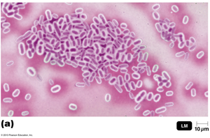
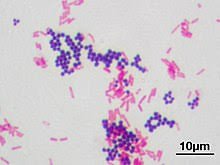
Q5. Why is the Gram stain so useful?
Background
Topic: Gram Staining and Bacterial Classification
This question tests your understanding of the Gram stain and its importance in differentiating bacterial types.
Key Terms and Formulas
Gram-positive: Bacteria with thick peptidoglycan cell walls, retain crystal violet stain.
Gram-negative: Bacteria with thin peptidoglycan and outer membrane, do not retain crystal violet.
Step-by-Step Guidance
Recall the steps of the Gram stain procedure.
Explain how the stain differentiates between Gram-positive and Gram-negative bacteria.
Discuss the clinical and diagnostic significance of Gram staining.
Try solving on your own before revealing the answer!
Final Answer:
The Gram stain is useful because it quickly differentiates bacteria into two major groups, guiding treatment and identification.
Q6. a. What characteristic of light do you know? b. How do you measure the wavelength? c. What unit is used to measure wavelength?
Background
Topic: Properties of Light in Microscopy
This question tests your understanding of light properties relevant to microscopy, especially wavelength.
Key Terms and Formulas
Wavelength (): The distance between two consecutive peaks of a wave.
Unit: Nanometers (nm)
Step-by-Step Guidance
Identify the main characteristics of light (e.g., wavelength, frequency, amplitude).
Describe how wavelength is measured (distance between peaks).
State the unit commonly used for wavelength in microscopy.
Try solving on your own before revealing the answer!
Final Answer:
Light is characterized by its wavelength, which is measured as the distance between two peaks and is typically expressed in nanometers (nm).
Analysis 1. In a Gram stain, a step could be omitted and still allow differentiation between gram-positive and gram-negative cells. What is it?
Background
Topic: Gram Staining Procedure
This question tests your knowledge of the steps in the Gram stain and which are essential for differentiation.
Key Terms and Formulas
Gram stain steps: Crystal violet, iodine, alcohol (decolorizer), safranin.
Step-by-Step Guidance
List the four main steps of the Gram stain.
Consider which steps are critical for differentiating Gram-positive and Gram-negative cells.
Think about which step could be omitted without losing the ability to distinguish the two groups.
Try solving on your own before revealing the answer!
Final Answer:
The final counterstain (safranin) can be omitted; Gram-positive cells will still appear purple, and Gram-negative cells will be colorless after decolorization.
Analysis 2. Using a good compound light microscope with a resolving power of 0.3 μm, a 10× ocular lens, and a 100× oil immersion lens, would you be able to discern two objects separated by 3 μm? 0.3 μm? 300 nm?
Background
Topic: Microscopy Resolution and Magnification
This question tests your understanding of resolving power and how it relates to the ability to distinguish objects at different distances.
Key Terms and Formulas
Resolving power: The minimum distance at which two points can be distinguished.
Magnification: Ocular lens × objective lens.
Step-by-Step Guidance
Calculate the total magnification: .
Compare the resolving power (0.3 μm) to the distances given (3 μm, 0.3 μm, 300 nm).
Determine which distances can be resolved based on the resolving power.
Try solving on your own before revealing the answer!
Final Answer:
Objects separated by 3 μm can be resolved; objects at 0.3 μm are at the limit; 300 nm (0.3 μm) is the minimum resolvable distance.
Analysis 4. Endospores can be seen as refractile structures in unstained cells and as colorless areas in Gram-stained cells. Why is it necessary to do an endospore stain to verify the presence of endospores?
Background
Topic: Endospore Staining
This question tests your understanding of why specific stains are needed to confirm the presence of endospores.
Key Terms and Formulas
Endospore: A resistant, dormant structure formed by some bacteria.
Endospore stain: Uses special dyes to specifically stain endospores.
Step-by-Step Guidance
Recall how endospores appear in unstained and Gram-stained preparations.
Explain why these appearances are not definitive for endospore identification.
Describe how the endospore stain provides confirmation.
Try solving on your own before revealing the answer!
Final Answer:
Endospore stains specifically color endospores, confirming their presence and distinguishing them from other structures.
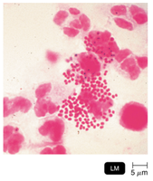
Clinical Application 2. Laboratory diagnosis of Neisseria gonorrhoeae infection is based on microscopic examination of Gram-stained pus. Locate the bacteria in this light micrograph. What is the disease?
Background
Topic: Clinical Microbiology and Gram Staining
This question tests your ability to interpret Gram-stained slides and relate findings to clinical diagnosis.
Key Terms and Formulas
Neisseria gonorrhoeae: Gram-negative diplococcus.
Gram stain: Used to visualize bacteria in clinical samples.
Step-by-Step Guidance
Examine the micrograph for small, Gram-negative (pink/red) diplococci.
Identify their location relative to host cells (often inside neutrophils).
Relate the findings to the clinical disease (gonorrhea).
Try solving on your own before revealing the answer!
Final Answer:
The bacteria are Gram-negative diplococci found within neutrophils; the disease is gonorrhea.
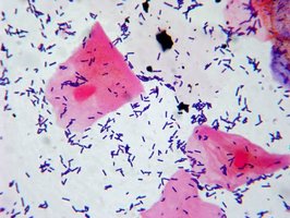
Clinical Application 3. Assume that you are viewing a Gram-stained sample of vaginal discharge. Large (10 μm) nucleated red cells are coated with small (0.5 μm wide by 1.5 μm long) blue cells on their surfaces. What is the most likely explanation for the red and blue cells?
Background
Topic: Clinical Microbiology and Gram Staining Interpretation
This question tests your ability to interpret Gram-stained slides and distinguish between host cells and bacteria.
Key Terms and Formulas
Gram stain: Differentiates cells based on cell wall structure.
Host cells: Large, nucleated (eukaryotic).
Bacterial cells: Small, blue (Gram-positive) or pink (Gram-negative).
Step-by-Step Guidance
Identify the large nucleated cells as host (human) cells.
Recognize the small blue cells as bacteria adhering to the host cells.
Consider the clinical significance of bacteria coating host cells.
Try solving on your own before revealing the answer!
Final Answer:
The red cells are host epithelial cells; the blue cells are Gram-positive bacteria adhering to them, indicating a bacterial infection.
Chapter 3 Clinical Case: List the 3 characteristics of Helicobacter pylori given in the case study:
Background
Topic: Clinical Microbiology Case Study
This question tests your ability to extract key characteristics of a pathogen from a clinical scenario.
Key Terms and Formulas
Helicobacter pylori: Gram-negative, spiral-shaped bacterium, causative agent of peptic ulcers.
Step-by-Step Guidance
Review the case study for descriptions of H. pylori's morphology, staining characteristics, and clinical relevance.
Identify three distinct characteristics mentioned in the case.
Relate these characteristics to laboratory identification methods.

Try solving on your own before revealing the answer!
Final Answer:
H. pylori is spiral-shaped, Gram-negative, and associated with peptic ulcers. Electron microscopy and Gram staining are used for identification.
