 Back
BackCharacterizing and Classifying Prokaryotes: Structure, Reproduction, and Diversity
Study Guide - Smart Notes
 Tailored notes based on your materials, expanded with key definitions, examples, and context.
General Characteristics of Prokaryotic Organisms
Diversity and Habitats
Prokaryotes are the most diverse group of cellular microbes, inhabiting a wide range of environments from extreme cold (Antarctic glaciers) to extreme heat (thermal hot springs), and from animal colons to the cytoplasm of other prokaryotes. They can also be found in distilled water, supersaturated brine, disinfectant solutions, and even basalt rocks. However, only a small fraction of prokaryotes are capable of colonizing humans and causing disease.
Typical Prokaryotic Morphologies
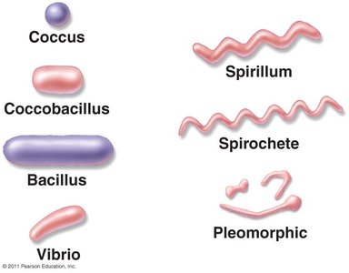
Prokaryotic cells exhibit a variety of shapes, which are important for classification and identification:
Coccus: Spherical-shaped cells
Bacillus: Rod-shaped cells
Coccobacillus: Short, oval rods resembling both cocci and bacilli
Vibrio: Comma-shaped cells
Spirillum: Rigid, spiral-shaped cells
Spirochete: Flexible, spiral-shaped cells
Pleomorphic: Cells that vary in shape and size
Reproduction of Prokaryotic Cells
Modes of Asexual Reproduction
All prokaryotes reproduce asexually, primarily through three main methods:
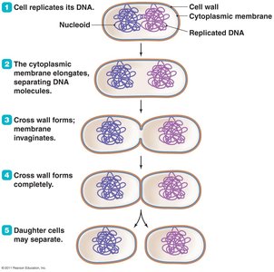
Binary Fission: The most common method, involving the replication of DNA, elongation of the cell, formation of a cross wall, and separation into two daughter cells.
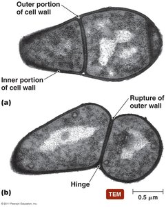
Snapping Division: A variation of binary fission where the inner portion of the cell wall forms a cross wall, but the outer wall ruptures, causing the cells to snap apart.
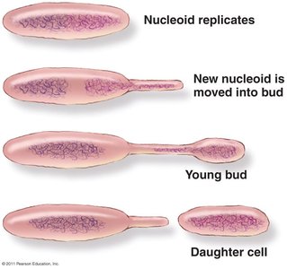
Budding: A process where a new cell develops from a bud due to the replication and migration of the nucleoid into the bud, which then separates as a daughter cell.
Binary Fission Process
Cell replicates its DNA.
The cytoplasmic membrane elongates, separating DNA molecules.
A cross wall forms and the membrane invaginates.
The cross wall completes formation.
Daughter cells may separate.
Snapping Division
Snapping division is characterized by the rupture of the outer cell wall, resulting in a hinge-like separation of daughter cells.
Budding
In budding, the nucleoid replicates and a new nucleoid is moved into a developing bud, which eventually separates as a new cell.
Other Reproductive Strategies
Viviparity: Some prokaryotes, such as Epulopiscium, reproduce by releasing live offspring from the body of the dead mother cell, a unique form of viviparous behavior in the prokaryotic world.
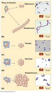
Arrangement of Prokaryotic Cells
Cellular Arrangements
The arrangement of prokaryotic cells is determined by the planes in which cells divide and whether daughter cells remain attached after division. Common arrangements include:
Cocci: Diplococci (pairs), Streptococci (chains), Tetrads (groups of four), Sarcinae (cuboidal packets), Staphylococci (clusters)
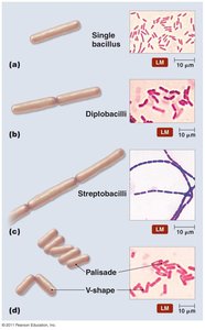
Bacilli: Single bacillus, Diplobacilli (pairs), Streptobacilli (chains), Palisade and V-shape arrangements
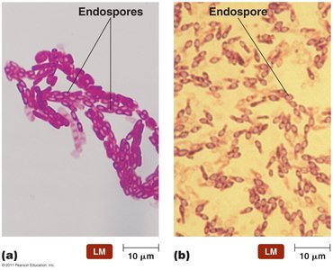
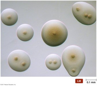
Endospores
Formation and Significance
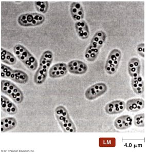
Endospores are highly resistant, dormant structures formed by certain Gram-positive bacteria such as Bacillus and Clostridium. Each vegetative cell transforms into one endospore, which can later germinate to form a new vegetative cell. Endospores serve as a defensive strategy against unfavorable environmental conditions and are of significant concern in food processing, healthcare, and government regulation due to their resistance to heat, desiccation, chemicals, and radiation.
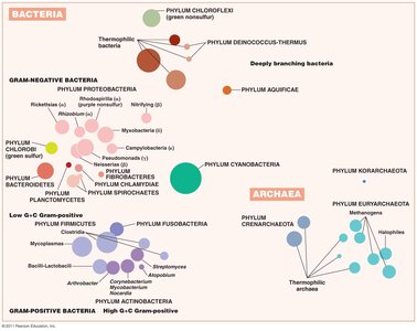
Modern Prokaryotic Classification
Taxonomic Domains
Modern classification of prokaryotes is based on genetic relatedness, particularly rRNA sequences. The three domains of life are:
Archaea
Bacteria
Eukarya
Survey of Archaea
General Features
Lack true peptidoglycan in their cell walls
Cell membrane lipids have branched hydrocarbon chains
The AUG codon codes for methionine (like eukaryotes)
Three main phyla: Crenarchaeota, Euryarchaeota, Korarchaeota
Reproduce by binary fission, budding, or fragmentation
Most are cocci, bacilli, or spiral forms; pleomorphic forms exist
Not known to cause disease in humans
Extremophiles
Extremophiles are archaea that require extreme conditions to survive, such as high temperature, extreme pH, or high salinity.
Thermophiles: Require temperatures above 45°C; hyperthermophiles require temperatures over 80°C. Examples include Geogemma and Pyrodictium.

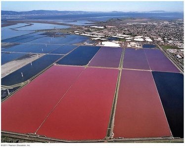
Halophiles: Inhabit highly saline environments, requiring more than 9% NaCl. Many produce red or orange pigments for protection against visible and UV light. The most studied is Halobacterium salinarium.
Methanogens
Methanogens are the largest group of archaea, producing methane gas from carbon dioxide, hydrogen gas, and organic acids. They play a crucial role in converting organic wastes in aquatic sediments to methane and are a primary source of environmental methane.
Survey of Bacteria
Deeply Branching and Phototrophic Bacteria
Deeply Branching Bacteria: Thought to resemble the earliest forms of bacteria; autotrophic and inhabit environments similar to early Earth. Examples include Aquifex and Deinococcus.
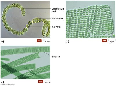
Phototrophic Bacteria: Contain photosynthetic lamellae and are divided into five groups based on pigments and electron sources: cyanobacteria (blue-green), green sulfur bacteria, green nonsulfur bacteria, purple sulfur bacteria, and purple nonsulfur bacteria.
Low G+C Gram-Positive Bacteria
Clostridia: Rod-shaped, obligate anaerobes important in medicine and industry.
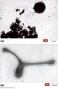
Mycoplasmas: Facultative or obligate anaerobes lacking cell walls; smallest free-living cells.
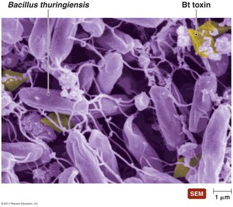
Bacillus: Common in soil; some species produce insecticidal toxins (e.g., Bacillus thuringiensis).
Listeria: Contaminates milk and meat products.
Lactobacillus: Grows in the body but rarely causes disease.
Streptococcus and Enterococcus: Cause numerous diseases.
Staphylococcus: Common inhabitants of humans.
High G+C Gram-Positive Bacteria
Corynebacterium: Pleomorphic aerobes and facultative anaerobes; produce metachromatic granules.
Mycobacterium: Aerobic rods with slow growth due to mycolic acid in cell walls.
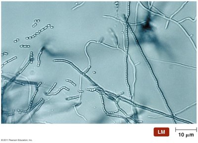
Actinomycetes: Form branching filaments resembling fungi; important genera include Actinomyces, Nocardia, and Streptomyces.
Gram-Negative Proteobacteria
Proteobacteria are the largest and most diverse group of bacteria, with many having prosthecae (extensions for attachment and nutrient absorption).
Alphaproteobacteria: Includes nitrogen fixers (Azospirillum, Rhizobium), nitrifying bacteria (Nitrobacter), purple nonsulfur phototrophs, and pathogens (Rickettsia, Brucella).
Betaproteobacteria: Includes pathogens (Neisseria, Bordetella, Burkholderia) and nonpathogens (Thiobacillus, Zoogloea, Sphaerotilus).
Gammaproteobacteria: Includes purple sulfur bacteria, intracellular pathogens (Legionella, Coxiella), methane oxidizers, glycolytic facultative anaerobes (family Enterobacteriaceae), and pseudomonads (Pseudomonas, Azotobacter, Azomonas).
Deltaproteobacteria: Includes Desulfovibrio, Bdellovibrio, and myxobacteria.
Epsilonproteobacteria: Includes Campylobacter and Helicobacter.
Other Gram-Negative Bacteria
Chlamydias: Genus Chlamydia
Spirochetes: Genera Treponema and Borrelia
Bacteroids: Genera Bacteroides and Cytophaga
Summary Table: Major Groups of Prokaryotes
Group | Key Features | Examples |
|---|---|---|
Archaea | No peptidoglycan, extremophiles, methanogens | Halobacterium, Geogemma |
Low G+C Gram-Positive | Low GC content, endospore formation, pathogens | Bacillus, Clostridium, Streptococcus |
High G+C Gram-Positive | High GC content, filamentous forms | Mycobacterium, Streptomyces |
Proteobacteria | Gram-negative, diverse metabolism | Rhizobium, Pseudomonas, Neisseria |
Other Gram-Negative | Unique cell structures, pathogens | Chlamydia, Borrelia |
Additional info: This summary table is inferred to provide a concise comparison of the major prokaryotic groups discussed in the notes.
