 Back
BackCharacterizing and Classifying Viruses, Viroids, and Prions
Study Guide - Smart Notes
 Tailored notes based on your materials, expanded with key definitions, examples, and context.
Characterizing and Classifying Viruses, Viroids, and Prions
Introduction
Viruses, viroids, and prions are acellular infectious agents that play significant roles in microbiology. They are responsible for numerous diseases in humans, animals, plants, and bacteria. Understanding their structure, replication, and classification is essential for microbiology students. 
Viruses
General Characteristics of Viruses
Viruses are minuscule infectious agents, typically measured in nanometers. They lack cellular structure and can possess either DNA or RNA, but never both. Viruses cannot carry out metabolic processes independently and must recruit host cell machinery for replication.
Acellular: No cytoplasmic membrane, cytosol, or organelles (with rare exceptions).
Extracellular State (Virion): Composed of a protein coat (capsid) surrounding nucleic acid; may have a phospholipid envelope.
Intracellular State: Capsid is removed, and the virus exists as nucleic acid within the host cell.
Diseases: Cause many infections and are responsible for most diseases in industrialized societies.
Structure of Virions
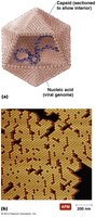
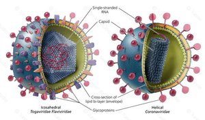
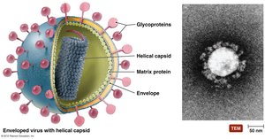
Virions consist of nucleic acid (DNA or RNA) encased in a protein coat called a capsid. Some viruses possess an envelope derived from the host cell membrane.
Nucleocapsid: Combination of nucleic acid and capsid.
Envelope: Phospholipid bilayer with glycoproteins, acquired during viral replication or release.
Recognition: Outermost layer provides protection and recognition sites for host cells.
Genetic Material of Viruses
The genetic material of viruses is the primary basis for their classification. Viral genomes may be DNA or RNA, single-stranded or double-stranded, and are much smaller than cellular genomes.
Types: dsDNA, ssDNA, dsRNA, ssRNA
Classification: Based on nucleic acid type and structure
Hosts and Specificity
Viruses exhibit specificity for their host cells, determined by the affinity of viral surface proteins for host cell receptors. Some viruses are highly specific, while others (generalists) can infect multiple cell types and hosts.
Host Range: Most viruses infect only particular cells in a specific host.
Generalist Viruses: Infect many kinds of cells in various hosts (e.g., rabies virus).
Size Comparison of Viruses
Viruses are much smaller than most cells and organelles, ranging from 24 nm to 300 nm. For example, poliovirus is 30 nm, while a red blood cell is 10,000 nm in diameter. 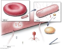
Viral Shapes
Viruses exhibit three basic shapes: helical, polyhedral, and complex. These shapes are determined by the arrangement of capsomeres in the capsid.
Helical: Rod-shaped, with nucleic acid spiraling inside.
Polyhedral: Many-sided, often icosahedral.
Complex: Combination of shapes, often seen in bacteriophages.
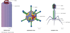
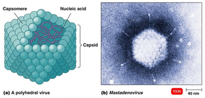
Capsid Morphology
Capsids protect viral nucleic acid and facilitate attachment to host cells. They are composed of protein subunits called capsomeres, which may be made of one or several types of proteins.
Protection: Shields nucleic acid from environmental damage.
Attachment: Enables binding to host cell receptors.
Viral Envelopes
Viral envelopes are acquired from the host cell membrane during replication or release. They consist of a phospholipid bilayer and proteins, including virally encoded glycoproteins (spikes) that play a role in host recognition.
Envelope Function: Facilitates entry into host cells and evasion of immune response.
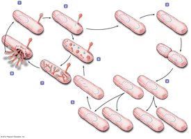
Viral Replication
Lytic Replication Cycle
The lytic cycle results in the destruction of the host cell and the release of new virions. It consists of five stages:
Attachment: Virus binds to host cell surface.
Entry: Viral genome enters the host cell.
Synthesis: Host machinery produces viral components.
Assembly: New virions are assembled.
Release: Host cell lyses, releasing virions.
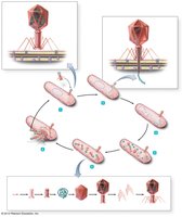
Lysogenic Replication Cycle
Lysogeny is a modified replication cycle in which the viral genome integrates into the host chromosome as a prophage. The host cell reproduces normally until induction triggers the lytic cycle.
Temperate Phages: Can switch between lysogenic and lytic cycles.
Lysogenic Conversion: Prophages may alter host phenotype.
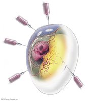
Replication of Animal Viruses
Animal viruses follow similar replication pathways as bacteriophages, with differences due to the presence of envelopes, eukaryotic cell structure, and lack of cell wall. Attachment is mediated by glycoprotein spikes or other molecules.
Attachment: Chemical attraction, no tails or tail fibers.
Entry: May occur via direct penetration, membrane fusion, or endocytosis.
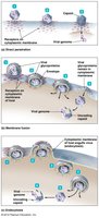
Synthesis and Release of Animal Viruses
The synthesis strategy depends on the type of nucleic acid. DNA viruses often replicate in the nucleus, while RNA viruses replicate in the cytoplasm. Enveloped viruses are released by budding, causing persistent infections; naked viruses are released by exocytosis or lysis.
Budding: Enveloped viruses exit the cell without immediate lysis.
Latency of Animal Viruses
Some animal viruses remain dormant in host cells for years, a state known as latency. Latent viruses may or may not integrate into the host genome; provirus incorporation is permanent.
Latent Infection: No viral activity for prolonged periods.
Role of Viruses in Cancer
Oncogene Theory
Viruses can induce cancer by disrupting normal cell division controls. They may carry oncogenes or interfere with tumor suppressor genes, leading to neoplasia (uncontrolled cell division).
Neoplasia: Formation of tumors; benign or malignant.
Malignant Tumors: Invade other tissues (metastasis).
Oncogene Activation: Environmental factors (UV, radiation, carcinogens, viruses) can activate oncogenes.
Viruses and Human Cancers
Viruses are implicated in 20-25% of human cancers. Examples include Burkitt’s lymphoma, Hodgkin’s disease, Kaposi’s sarcoma, and cervical cancer.
Mechanisms: Carry oncogenes, promote host oncogenes, or interfere with tumor repression.
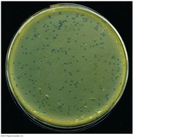
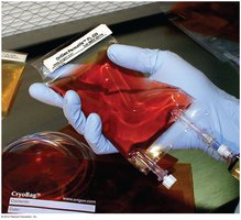
Culturing Viruses
Methods of Culturing
Viruses can be cultured using mature organisms (bacteria, plants, animals), embryonated chicken eggs, or cell (tissue) cultures. Cell cultures are classified as diploid or continuous.
Bacterial Lawn: Used to observe viral plaques.
Embryonated Eggs: Provide a sterile, nutrient-rich environment.
Cell Cultures: Isolated cells grown in media; diploid and continuous types.
Viroids
Characteristics of Viroids
Viroids are extremely small, circular RNA molecules that infect plants. They lack a capsid and membrane, distinguishing them from viruses. Viroids may appear linear due to hydrogen bonding.
Pathogenicity: Cause plant diseases.
Structure: Similar to RNA viruses but simpler.
Prions
Characteristics of Prions
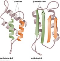
Prions are infectious proteins that cause fatal neurological diseases. They are misfolded forms of normal cellular PrP protein, with beta-pleated sheets instead of alpha-helices. Prions induce misfolding in normal proteins, leading to disease.
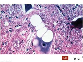
Prion Diseases: Spongiform encephalopathies, characterized by vacuole formation and loss of brain matter.
Destruction: Prions are resistant to most methods; destroyed by incineration or autoclaving in 1 M NaOH.
Are Viruses Alive?
Debate on Viral Life
There is ongoing debate about whether viruses are living entities. Some consider them complex pathogenic chemicals, while others view them as the simplest forms of life due to their ability to invade cells and replicate.
Arguments for Life: Ability to replicate and control host cells.
Arguments against Life: Lack of independent metabolism and cellular structure.
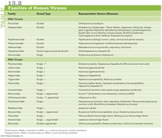
Summary Table: Families of Human Viruses
Classification by Genome Type
Viruses are classified into families based on their genome type (DNA or RNA), strand type, and representative genera/diseases.
Family | Strand Type | Representative Genera/Diseases |
|---|---|---|
DNA Viruses | Double/Single | Orthopoxvirus (smallpox), Herpesvirus (herpes, chickenpox), Papillomavirus (warts), Adenovirus (respiratory infections) |
RNA Viruses | Single/Double | Enterovirus (polio), Flavivirus (yellow fever), Orthomyxovirus (influenza), Retrovirus (HIV/AIDS) |
Additional info: | Families and diseases inferred from typical textbook tables | See textbook for full list |
 ----------------------------------------
