 Back
BackCharacterizing and Classifying Viruses, Viroids, and Prions: Study Notes
Study Guide - Smart Notes
 Tailored notes based on your materials, expanded with key definitions, examples, and context.
Characterizing and Classifying Viruses, Viroids, and Prions
Viral Replication in Animal Cells
Animal viruses replicate using pathways similar to bacteriophages, but with distinct differences due to the presence of envelopes, the eukaryotic nature of animal cells, and the absence of cell walls. The replication process involves several stages: attachment, entry, uncoating, synthesis, assembly, and release.
Attachment
Chemical attraction occurs between viral proteins and cell receptors.
Animal viruses lack tails or tail fibers; instead, they use glycoprotein spikes or other attachment molecules.
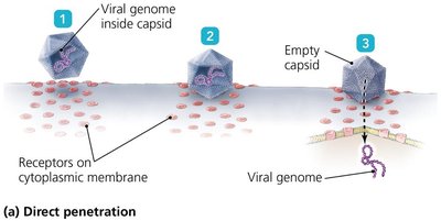
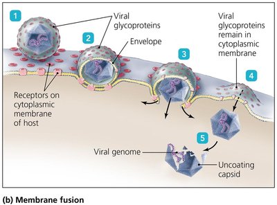
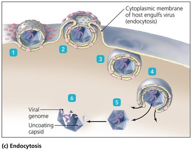
Entry and Uncoating
Viruses enter host cells by three main mechanisms:
Direct penetration: Viral genome enters the cell, leaving the capsid outside.
Membrane fusion: Viral envelope fuses with the host membrane, releasing the genome.
Endocytosis: Host cell engulfs the virus, which is then uncoated inside the cell.
Synthesis
DNA viruses often enter the nucleus for replication.
RNA viruses typically replicate in the cytoplasm.
Retroviruses use a DNA intermediary transcribed by viral reverse transcriptase.
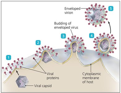
Assembly and Release
Most DNA viruses assemble in the nucleus; most RNA viruses assemble in the cytoplasm.
Enveloped viruses acquire host membrane during release (budding).
Naked viruses are released by exocytosis or lysis.
Latency
Some animal viruses remain dormant as latent viruses or proviruses.
Latency may last years; provirus incorporation into host DNA is permanent.
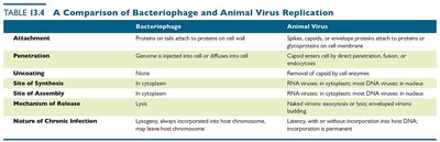
Comparison of Bacteriophage and Animal Virus Replication
This table summarizes the key differences between bacteriophage and animal virus replication.
Step | Bacteriophage | Animal Virus |
|---|---|---|
Attachment | Proteins on tail attach to proteins on cell wall | Spikes, capsid, or envelope proteins attach to proteins or glycoproteins on cell membrane |
Penetration | Genome is injected into cell or diffuses into cell | Capsid enters cell by direct penetration, fusion, or endocytosis |
Uncoating | None | Removed by cellular enzymes |
Site of Synthesis | In cytoplasm | RNA viruses in cytoplasm; most DNA viruses in nucleus |
Site of Assembly | In cytoplasm | RNA viruses in cytoplasm; most DNA viruses in nucleus |
Mechanism of Release | Lysis | Budding (enveloped viruses) or exocytosis/lysis (naked viruses) |
Nature of Chronic Infection | Lysogeny; always incorporated into host chromosome, may leave host chromosome | Latency, with or without incorporation into host DNA; incorporation is permanent |
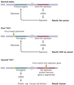
The Role of Viruses in Cancer
Viruses can contribute to cancer by affecting protooncogenes and oncogenes, which regulate cell growth and division. Environmental factors and viral infections can activate oncogenes, leading to uncontrolled cell division.
Viruses may carry oncogenes or promote activation of host oncogenes.
Some viruses interfere with tumor repression.
Examples of virus-induced cancers: Burkitt’s lymphoma, Hodgkin’s disease, Kaposi’s sarcoma, cervical cancer.
Culturing Viruses in the Laboratory
Viruses require host cells for growth and cannot be cultured in standard microbiological media. Three main methods are used: mature organisms, embryonated eggs, and cell cultures.
Culturing in Mature Organisms
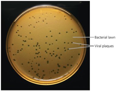
Bacteriophages are grown in bacteria using liquid cultures or agar plates.
Lysis of bacteria produces plaques, which allow estimation of phage numbers by plaque assay.
Culturing in Plants and Animals
Viruses can be cultured in various plants and animals, though laboratory animals are costly and raise ethical concerns.
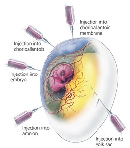
Culturing in Embryonated Chicken Eggs
Chicken eggs are inexpensive, large, free of contaminants, and contain nourishing yolk.
Embryonic tissues provide ideal sites for viral growth; some vaccines are prepared in chicken cultures.
Other Parasitic Particles: Viroids and Prions
Viroids
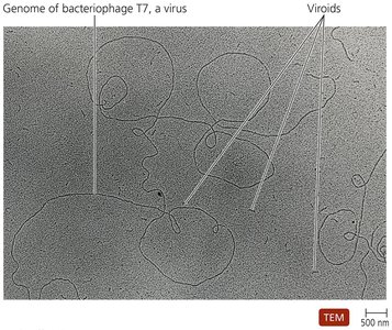
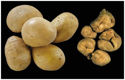
Viroids are extremely small, circular pieces of single-stranded RNA (ssRNA) that are infectious and pathogenic in plants. They lack a capsid and do not code for proteins.
Viroid RNA adheres to complementary plant RNA, leading to degradation by plant enzymes and disease.
Prions
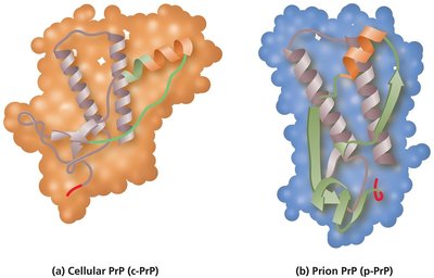
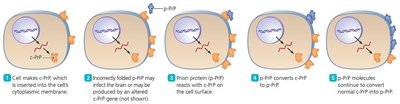
Prions are proteinaceous infectious agents. They exist in two forms:
Cellular PrP: Normal, functional structure with α-helices.
Prion PrP: Disease-causing form with β-pleated sheets.
Prion PrP induces cellular PrP to refold into the prion form, leading to disease.
Prion Diseases
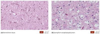
Prion diseases include spongiform encephalopathies (e.g., BSE, scrapie, kuru, CWD, vCJD).
Characterized by large vacuoles in brain tissue, giving a spongy appearance.
Transmitted by ingestion, transplantation, or contact with infected tissues.
No standard treatment; prions are resistant to normal sterilization, destroyed by incineration or autoclaving in concentrated sodium hydroxide.
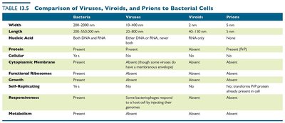
Comparison of Viruses, Viroids, Prions, and Bacterial Cells
This table compares the main properties of bacteria, viruses, viroids, and prions.
Property | Bacteria | Viruses | Viroids | Prions |
|---|---|---|---|---|
Width | 200–2000 nm | 10–400 nm | 2–10 nm | 5 nm |
Length | 200–550,000 nm | 10–400 nm | 2–10 nm | 5 nm |
Nucleic Acid | Both DNA and RNA | Either DNA or RNA, never both | RNA only | None |
Protein | Present | Present | Absent | Present |
Cellular | Yes | No | No | No |
Cytoplasmic Membrane | Present | Absent (some viruses have a membranous envelope) | Absent | Absent |
Functional Ribosomes | Present | Absent | Absent | Absent |
Growth | Yes | No | No | No |
Self-Replicating | Yes | No | No | No, transforms PrP protein present in cell |
Responsiveness | Present | Some bacteriophages respond to a host cell by injecting their genome | Absent | Absent |
Metabolism | Present | Absent | Absent | Absent |
Additional info: The notes above expand on brief points with academic context, definitions, and examples to ensure completeness and clarity for microbiology students.
