 Back
BackCharacterizing and Classifying Viruses, Viroids, and Prions
Study Guide - Smart Notes
 Tailored notes based on your materials, expanded with key definitions, examples, and context.
Characterizing and Classifying Viruses, Viroids, and Prions
Introduction to Acellular Infectious Agents
Viruses, viroids, and prions are acellular agents responsible for a wide range of diseases in humans, animals, plants, and bacteria. Unlike cellular life forms, these agents lack the ability to carry out metabolic processes independently and must exploit host cells for replication and propagation.
Viruses: Minuscule, acellular infectious agents containing either DNA or RNA, but never both.
Viroids: Infectious RNA molecules, primarily affecting plants.
Prions: Infectious proteins lacking nucleic acids, causing neurodegenerative diseases.
These agents are obligate intracellular parasites, meaning they require host cell machinery for replication.
Characteristics of Viruses
General Properties
Viruses are distinguished by their inability to perform metabolic activities, grow, or respond to the environment outside a host cell. They exist in two states:
Extracellular state (Virion): Composed of a nucleic acid core surrounded by a protein coat (capsid), and sometimes a phospholipid envelope.
Intracellular state: The capsid is removed, and the virus exists as nucleic acid within the host cell.
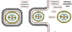
The outermost layer of the virion provides protection and recognition sites for host cells.
Distinguishing Features of Viruses
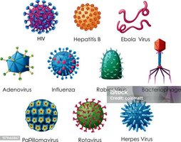
Type of genetic material (DNA or RNA; single or double-stranded)
Kinds of cells they infect (host range)
Size and shape
Nature of the capsid coat
Presence or absence of an envelope
Genetic Material of Viruses
Viral genomes are highly variable and are the primary basis for classification. They may be:
DNA or RNA (never both)
Single-stranded (ss) or double-stranded (ds)
Linear or circular
Viral genomes are much smaller than those of cells, with some viruses encoding as few as three genes.
Host Range and Specificity
Most viruses infect specific cell types due to the affinity between viral surface proteins and host cell receptors. Some, called generalists, can infect multiple cell types or species.
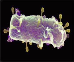
Bacteriophages: Viruses that infect bacteria.
All life forms are susceptible to viral infection.
Size of Viruses
Viruses are extremely small, typically ranging from 10 nm to 300 nm, making them invisible under light microscopy.
Capsid Morphology and Viral Shapes
Capsid Structure
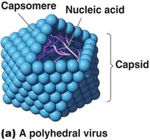
The capsid is a protein shell composed of subunits called capsomeres. It protects the viral genome and facilitates attachment to host cells.
Viral Shapes
Viruses exhibit three basic shapes:
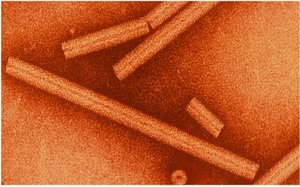
Helical: Capsomeres spiral around the nucleic acid, forming a tube-like structure.
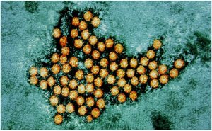
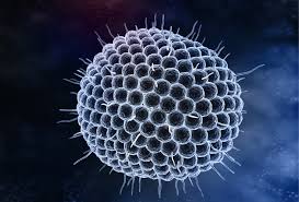
Polyhedral: Roughly spherical, resembling a geodesic dome.
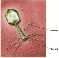
Complex: Capsids with intricate structures, often seen in bacteriophages.
Viral Envelopes
Structure and Function
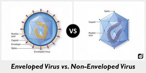
Some viruses possess an envelope derived from the host cell membrane, which surrounds the capsid. Enveloped viruses are distinct from non-enveloped (naked) viruses.
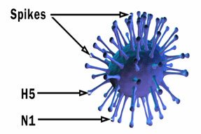
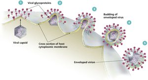
The envelope consists of a phospholipid bilayer and proteins, including virally encoded glycoproteins (spikes).
Envelope proteins play a crucial role in host cell recognition and attachment.
Classification of Viruses
Taxonomy
The International Committee on Taxonomy of Viruses (ICTV) classifies viruses based on genetic material, morphology, and replication strategy. The highest recognized taxa are orders, followed by families and genera. Species names are typically descriptive (e.g., Rabies virus, Herpes virus).
Viral Replication
Lytic Replication Cycle
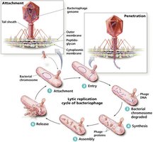
The lytic cycle is a common replication strategy for bacteriophages, resulting in the destruction of the host cell and release of new virions. The stages include:
Attachment
Entry
Synthesis
Assembly
Release

Lysogenic Replication Cycle
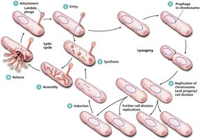
Some bacteriophages can integrate their genome into the host chromosome, becoming a prophage. This lysogenic cycle allows the viral genome to be replicated along with the host cell's DNA until induction triggers entry into the lytic cycle.
Animal Virus Replication
Attachment and Entry
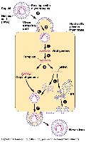
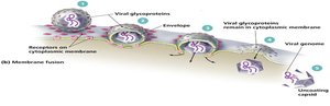
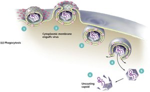
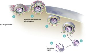
Animal viruses attach to host cells via glycoprotein spikes or other molecules. Entry mechanisms include direct penetration, membrane fusion, and endocytosis (phagocytosis).
Specificity of attachment determines host range (e.g., human vs. avian influenza).

Synthesis of Viral Components
The strategy for viral genome replication and protein synthesis depends on the type of nucleic acid:
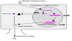
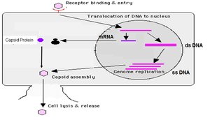
dsDNA viruses: Replicate in the nucleus using host enzymes.
ssDNA viruses: Synthesize a complementary strand to form dsDNA, then proceed as dsDNA viruses.
RNA viruses: Use various strategies depending on whether the RNA is positive-sense, negative-sense, or double-stranded.
RNA Virus Replication Strategies
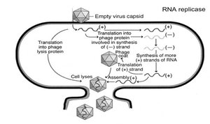
+ssRNA viruses: Genome acts as mRNA for direct translation.
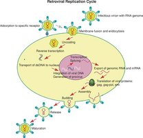
Retroviruses: +ssRNA is reverse transcribed into DNA, which integrates into the host genome.
-ssRNA viruses: Carry RNA-dependent RNA transcriptase to synthesize mRNA from their genome.
dsRNA viruses: Each strand serves as a template for replication and protein synthesis.

Assembly and Release
New virions are assembled spontaneously. Enveloped viruses are typically released by budding, acquiring their envelope from the host cell membrane, which allows the host cell to survive longer than in lytic release.
Latency in Animal Viruses
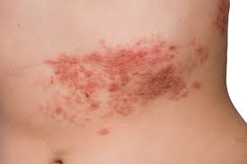
Some animal viruses can remain dormant within host cells, a state known as latency. Latent viruses may integrate into the host genome (provirus) or persist as episomes. Reactivation can occur years later, as seen in herpesviruses (e.g., chickenpox and shingles).

Viruses and Cancer
Oncogenesis
Viruses can contribute to cancer development by disrupting normal regulation of cell division. This may involve activation of proto-oncogenes or inactivation of tumor suppressor genes, leading to uncontrolled cell proliferation (neoplasia).

Culturing Viruses
Laboratory Methods
Because viruses require living cells for replication, they are cultured in whole organisms (bacteria, plants, animals), embryonated eggs, or cell (tissue) cultures.

Viroids and Prions
Viroids
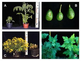
Viroids are small, circular RNA molecules that infect plants, causing diseases in crops such as potatoes, avocados, and coconuts. They lack a protein coat and are similar to RNA viruses but are smaller and simpler.
Prions
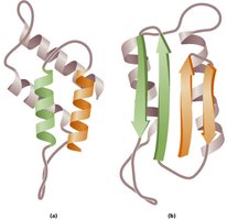
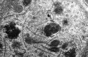
Prions are infectious proteins that lack nucleic acids. They cause fatal neurodegenerative diseases by inducing abnormal folding of normal cellular proteins (PrP) into a disease-causing form. Prion diseases are characterized by spongiform changes in brain tissue and are resistant to standard sterilization methods.
Examples: Bovine spongiform encephalopathy (mad cow disease), scrapie, Creutzfeldt-Jakob disease, Kuru.
Are Viruses Alive?
The debate over whether viruses are alive centers on their lack of independent metabolism and cellular structure. Outside host cells, viruses are inert; inside, they direct the synthesis and assembly of new virions, blurring the line between living and non-living entities.
