 Back
Characterizing and Classifying Viruses, Viroids, and Prions
Study Guide - Smart Notes
 Tailored notes based on your materials, expanded with key definitions, examples, and context.
General Characteristics of Viruses
Alive? The Nature of Viruses
Viruses are unique biological entities that blur the line between living and non-living. They are obligate intracellular parasites, meaning they require a host cell to reproduce and cannot carry out metabolic processes independently.
Genetic Material: Viruses contain either DNA or RNA, but never both.
Protein Coat: Their genetic material is surrounded by a capsid (protein coat).
Replication: Viruses hijack the host cell's machinery to multiply, as they lack their own enzymes for metabolism.
Transfer Structures: They synthesize structures to transfer their nucleic acid to other cells.
Why can’t an antiviral drug inhibit viral multiplication? Most antiviral drugs target viral enzymes or processes, but viruses rely almost entirely on host cell machinery, making selective inhibition difficult.
Characteristics of Viruses
Extracellular and Intracellular States
Viruses exist in two distinct states:
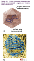
Extracellular (Virion): The virus is outside a host cell, with a capsid surrounding its nucleic acid. Some have a phospholipid envelope.
Intracellular: The capsid is removed, and the virus exists as naked nucleic acid within the host cell.
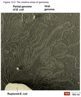
Structure of Viral Genomes
Nucleic Acid Types
Viral genomes are highly variable:
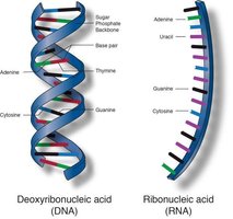
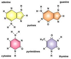
Can be DNA or RNA (never both).
May be single-stranded or double-stranded.
Can be linear or circular.
Structure: Capsid and Envelope
Capsid and Envelope Components
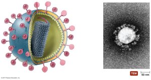
Capsid: Protein coat composed of subunits called capsomeres, protecting viral nucleic acid.
Envelope: Some viruses acquire a phospholipid envelope from the host cell during replication or release, often with glycoprotein spikes.
Mutability: Envelope proteins are highly mutable, affecting antibody and vaccine effectiveness.
Viral Morphology
Shapes and Structures
Viruses display diverse morphologies:
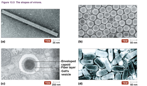
Helical: Cylindrical capsid with nucleic acid inside.
Polyhedral: Many-sided, often icosahedral (20 faces).
Enveloped: Spherical, with helical or polyhedral capsid inside.
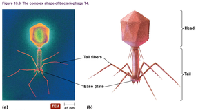
Complex: Capsids with additional structures, such as tail fibers.
Host Range and Specificity
Host Range
Viruses can infect all forms of life, but many are specific to certain cell types or species. This specificity is determined by:
Attachment Capabilities: Ability to bind to host cell receptors.
Machinery Compatibility: Host cell must support viral replication.
Bacteriophages (phages) infect bacteria and are used in research and therapeutic applications.
Size Comparison of Virions
Relative Sizes
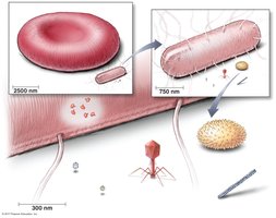
Viruses are much smaller than cells and organelles.
Red blood cell: 10,000 nm
E. coli: 1,000 x 3,000 nm
Bacterial ribosome: 25 nm
Poliovirus: 30 nm
Bacteriophage MS2: 24 nm
Bacteriophage T4: 50 x 225 nm
Smallpox virus: 200 x 300 nm
Tobacco mosaic virus: 15 x 300 nm
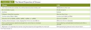
Novel Properties of Viruses
Comparison to Cells
Viruses differ fundamentally from cells in several ways:
Viruses | Cells |
|---|---|
Inert outside host cell | Metabolize independently |
Do not divide or grow | Divide and grow |
Obligate intracellular parasites | Cellular |
Contain either DNA or RNA | Contain both DNA and RNA |
Highly diverse in size (10–500 nm) | 200 nm to 12 cm |
Protein coat (capsid) | Surrounded by plasma membrane |
Replicate using host enzymes | Self-replicating by mitosis/meiosis |
Classification of Viruses
Criteria for Classification
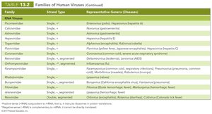
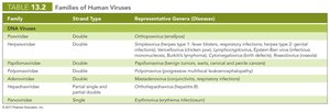
Virus classification is based on:
Type of nucleic acid (DNA or RNA)
Presence of an envelope
Shape
Size
Viral genera are organized into families; relationships among viruses are not fully understood.
Viral Replication in Bacteria
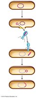
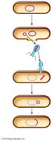
Lytic and Lysogenic Cycles
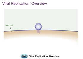
Viral replication in bacteria occurs via two main cycles:
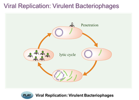
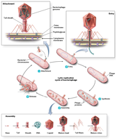
Lytic Cycle: Results in host cell lysis and death.
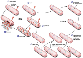
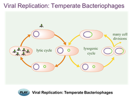
Lysogenic Cycle: Viral DNA integrates into host genome and remains latent.
Five stages of lytic cycle: Attachment, Entry, Synthesis, Assembly, Release.
Lysogenic Cycle
Phage DNA integrates into host DNA (prophage).
Host cell divides, passing prophage to daughter cells.
Prophage may excise and enter lytic cycle.
Consequences of Lysogeny
Lysogenic cells are immune to infection by same phage type.
Phage conversion: Host cell acquires new properties (e.g., toxin production).
Specialized transduction: Both phage and host DNA are transferred to new host.
Multiplication of Animal Viruses
Attachment and Entry
Attachment: Chemical attraction between viral proteins and cell receptors.
Entry: Three mechanisms—direct penetration, receptor-mediated endocytosis, fusion (for enveloped viruses).
Uncoating: Capsid is removed, releasing viral genome.
Eclipse period: Virus is disassembled and not detectable.
Synthesis, Maturation, and Release
DNA Viruses: DNA replicated in nucleus; proteins synthesized in cytoplasm.
RNA Viruses: RNA replicated in cytoplasm; positive-sense RNA acts as mRNA, negative-sense RNA must be transcribed.
Release: Enveloped viruses bud from cell; non-enveloped viruses cause cell rupture.
Retroviruses
Contain reverse transcriptase enzyme.
Copy viral RNA to produce double-stranded DNA, which integrates into host genome as a provirus.
Provirus is permanent and cannot be excised.
Latent vs Persistent Viral Infections
Types of Infection
Latent Infection: Virus remains dormant in host cell for long periods without causing symptoms.
Persistent Infection: Disease develops gradually and is often fatal.
The Role of Viruses in Cancer
Oncogenes and Tumor Formation
Protooncogenes: Promote cell growth and division.
Uncontrolled activation leads to cancer.
Some viruses carry oncogenes or activate host oncogenes.
Viruses cause 20–25% of human cancers (e.g., Burkitt's lymphoma, Hodgkin's disease, Kaposi's sarcoma, cervical cancer).
Culturing Viruses in the Laboratory
Methods of Culturing
Viruses require host cells for growth.
Three types of media: mature organisms, embryonated eggs, cell cultures.
Bacteriophages: Plaque method used to detect and count viruses (measured in plaque-forming units, PFU).
Culturing Animal Viruses
Living animals, embryonated eggs, and cell cultures are used.
Cell lines: primary, diploid, continuous (immortal).
Cytopathic effect (CPE) is measured to assess viral growth.
Identification of Viruses
Methods
Electron microscopy for visualization.
Serological methods (antibody detection).
Cytopathic effects.
RFLP and PCR for DNA amplification.
Plant Viruses and Viroids
Plant Virus Effects
Cause color change, deformed growth, wilting, stunted growth.
Infection often requires cell wall damage (parasites, wounds, insect feeding).
Some plant viruses multiply within insects and spread via pollen.
Viroids
Extremely small, circular pieces of single-stranded RNA.
Infectious and pathogenic in plants.
Lack capsid; do not code for proteins.
Viroid RNA adheres to complementary plant RNA, leading to degradation and disease.
Other Parasitic Particles: Prions
Characteristics of Prions
Proteinaceous infectious agents.
Can be destroyed by proteases or incineration.
Alter normal proteins into infectious forms.
Cause neurological diseases (spongiform encephalopathies): Mad Cow disease, Creutzfeldt-Jakob, scrapie, kuru.
Transmitted by ingestion, transplantation, contact with infected tissues, or genetically.
No standard treatment for prion diseases.
Comparison of Viruses, Viroids, Prions, and Bacterial Cells
Key Differences
Entity | Genetic Material | Structure | Replication | Pathogenicity |
|---|---|---|---|---|
Virus | DNA or RNA | Capsid, sometimes envelope | Host cell machinery | Animals, plants, bacteria |
Viroid | ssRNA | No capsid | Host cell enzymes | Plants |
Prion | None | Protein only | Induces misfolding | Animals (neurological) |
Bacterial Cell | DNA | Cell wall, membrane | Binary fission | Animals, plants |
