 Back
BackCharacterizing and Classifying Viruses, Viroids, and Prions: Study Notes
Study Guide - Smart Notes
 Tailored notes based on your materials, expanded with key definitions, examples, and context.
Characterizing and Classifying Viruses, Viroids, and Prions
Characteristics of Viruses
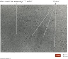
Viruses are minuscule, acellular infectious agents that possess either DNA or RNA as their genetic material. They are responsible for numerous diseases affecting humans, animals, plants, and bacteria. Unlike cellular organisms, viruses cannot carry out metabolic processes, grow, or respond to their environment independently. Instead, they rely on host cells to reproduce and increase their numbers. Viruses lack cytoplasmic membranes, cytosol, and organelles, and exist in two distinct states: extracellular and intracellular.
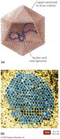
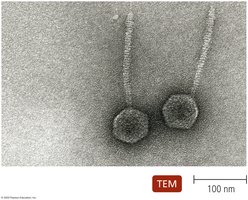
Extracellular State (Virion): The virion consists of a protein coat (capsid) surrounding the nucleic acid, forming the nucleocapsid. Some virions possess a phospholipid envelope, which provides protection and recognition sites for host cells.
Intracellular State: The capsid is removed, and the virus exists as nucleic acid within the host cell.
Genetic Material of Viruses
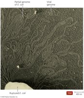
Viruses exhibit remarkable diversity in their genomes, which is a primary basis for their classification. Their genetic material may be DNA or RNA, but never both. Viral genomes can be double-stranded (dsDNA, dsRNA) or single-stranded (ssDNA, ssRNA), linear and segmented, or single and circular. Viral genomes are much smaller than those of cells.
Hosts of Viruses
Most viruses infect only specific host cells due to the affinity between viral surface proteins and complementary proteins on the host cell surface. Some viruses are highly specific, infecting only a particular cell type in a particular host, while others are generalists, infecting multiple cell types or hosts. All types of organisms are susceptible to viral infection.

Capsid Morphology and Viral Shapes
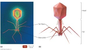
The capsid provides protection for the viral nucleic acid and facilitates attachment to host cells. Capsids are composed of protein subunits called capsomeres, which may consist of one or several types of proteins. Viruses are classified by their virion shape, with three basic types:
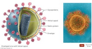
Helical
Polyhedral
Complex

The Viral Envelope
Some viruses acquire an envelope from the host cell during replication or release. The envelope is a portion of the host's membrane system, composed of a phospholipid bilayer and proteins, including virally coded glycoproteins (spikes). These proteins play a crucial role in host recognition. Enveloped viruses are generally more fragile than naked viruses.
Classification of Viruses
Viruses are classified based on the type of nucleic acid, presence of an envelope, shape, and size. Viral genera are organized into families, but relationships among viruses are not fully understood by taxonomists.
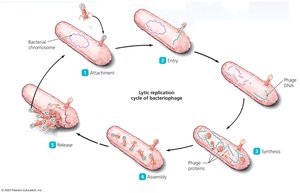
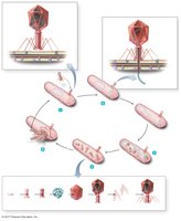
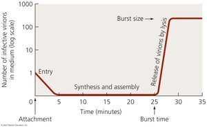
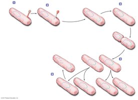
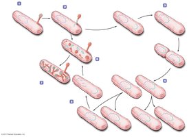
Viral Replication: Lytic Cycle
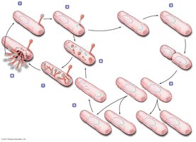
Viral replication depends on host organelles and enzymes. The lytic replication cycle typically results in the death and lysis of the host cell and consists of five stages:
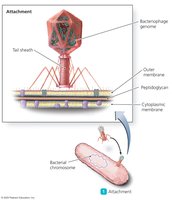
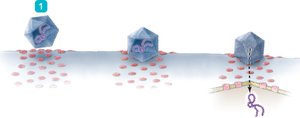
Attachment
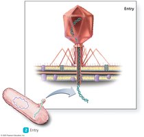
Entry
Synthesis
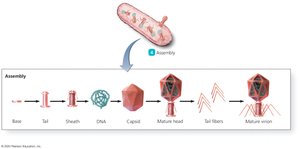
Assembly
Release
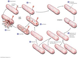
Viral Replication: Lysogenic Cycle
The lysogenic replication cycle is a modified process in which infected host cells grow and reproduce normally for generations before lysing. This occurs with temperate phages, which are called prophages during their inactive state. Lysogenic conversion can result when phages carry genes that alter the phenotype of a bacterium.


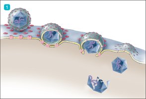
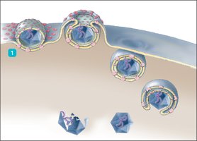
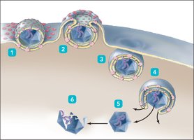
Replication of Animal Viruses
Animal viruses follow a similar basic replication pathway as bacteriophages, but differences arise due to the presence of an envelope, the eukaryotic nature of animal cells, and the lack of a cell wall. Attachment occurs via chemical attraction between viral proteins and cell receptors, often mediated by glycoprotein spikes. Entry and uncoating can occur by direct penetration, membrane fusion, or endocytosis.
Direct Penetration: Viral genome enters the cell directly.
Membrane Fusion: Viral envelope fuses with the host membrane, releasing the genome.
Endocytosis: Host cell engulfs the virus.
Synthesis of Animal Viruses
The synthesis strategy depends on the type of viral nucleic acid:
dsDNA Viruses: Replicate genome in the nucleus; proteins are synthesized in the cytoplasm.
ssDNA Viruses: ssDNA folds back to form dsDNA, which is replicated by cellular DNA polymerase.
RNA Viruses: Replication occurs in the cytoplasm. Positive-sense (+) RNA acts as mRNA; negative-sense (−) RNA must be transcribed to mRNA.
Retroviruses: Use a DNA intermediary transcribed by reverse transcriptase.

Assembly and Release of Animal Viruses
Most DNA viruses assemble in the nucleus, while most RNA viruses assemble in the cytoplasm. Enveloped viruses are released via budding, which can result in persistent infections. Naked viruses are released by exocytosis or lysis.
The Role of Viruses in Cancer
Cell division is tightly regulated by genetic controls. Neoplasia refers to uncontrolled cell division, resulting in tumors, which may be benign or malignant (cancer). Viruses may cause 20–25% of human cancers by carrying oncogenes, promoting host oncogenes, or interfering with tumor repression. Environmental factors such as UV light, radiation, carcinogens, and viruses can activate oncogenes.
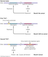
Examples of virus-induced cancers: Burkitt’s lymphoma, Hodgkin’s disease, Kaposi’s sarcoma, cervical cancer.
Other Parasitic Particles: Viroids and Prions
Viroids
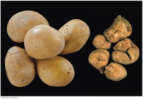
Viroids are extremely small, circular pieces of ssRNA that are infectious and pathogenic in plants. They lack a capsid and do not code for proteins. Viroid RNA adheres to complementary plant RNA, leading to degradation by plant enzymes and resulting in disease.
Prions
Prions are proteinaceous infectious agents. Normal cellular PrP has α-helices, while disease-causing prion PrP has β-pleated sheets. Prion PrP induces refolding of cellular PrP into the prion form, leading to spongiform encephalopathies characterized by large vacuoles in the brain. Prion diseases include BSE, scrapie, kuru, CWD, and vCJD. Prions are resistant to standard sterilization and are destroyed by incineration or autoclaving in concentrated sodium hydroxide.
Comparison Table: Viruses, Viroids, Prions, and Bacterial Cells
Property | Viruses | Viroids | Prions | Bacterial Cells |
|---|---|---|---|---|
Genetic Material | DNA or RNA | ssRNA | None | DNA |
Capsid | Present | Absent | Absent | Absent |
Envelope | Sometimes | Absent | Absent | Absent |
Replication | Host cell machinery | Host cell machinery | Induces misfolding | Binary fission |
Diseases | Various | Plant diseases | Spongiform encephalopathies | Various |
Size | Nanometers | Nanometers | Nanometers | Micrometers |
Additional info: | Viruses may be enveloped or naked | Viroids affect only plants | Prions are proteins only | Bacteria are cellular organisms |
