Back
Backchapter 13
Study Guide - Smart Notes
 Tailored notes based on your materials, expanded with key definitions, examples, and context.
Characterizing and Classifying Viruses, Viroids, and Prions
Are Viruses Alive?
Viruses occupy a unique position at the boundary between living and nonliving entities. Some microbiologists consider them complex pathogenic chemicals, while others view them as the least complex living entities. Viruses use sophisticated mechanisms to invade host cells, commandeer cellular machinery, and replicate their genomes. However, they lack independent metabolic activity and cannot reproduce outside a host cell.
Pathogenic chemicals: Viruses do not carry out metabolic processes independently.
Living entities: They possess genomes and can direct their own replication within host cells.
Characteristics of Viruses
Viruses are minuscule, acellular infectious agents that contain either DNA or RNA as their genetic material. They infect a wide range of hosts, including humans, animals, plants, and bacteria, and are responsible for many significant diseases. Viruses lack cellular structures such as cytoplasmic membranes, cytosol, and organelles, and cannot grow, respond to the environment, or reproduce independently.
Extracellular state (virion): Composed of a protein coat (capsid) surrounding nucleic acid; may have a phospholipid envelope.
Intracellular state: Capsid is removed, and the virus exists as nucleic acid within the host cell.
Comparison of Viruses and Cells
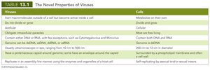
Viruses differ fundamentally from cells in structure, replication, and function. The following table summarizes the novel properties of viruses compared to cells:
Viruses | Cells |
|---|---|
Obligate intracellular parasites | May live free-living |
Contain either DNA or RNA, never both | Contain both DNA and RNA |
Acellular | Cellular |
Do not divide or grow | Divide and grow |
Replicate in an assembly-line manner using host enzymes | Self-replicating by asexual and/or sexual means |
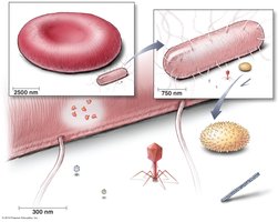
Usually submicroscopic (20–300 nm) | 200 nm to several centimeters in diameter |
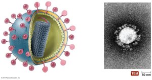
Structure of Virions
A complete virus particle, or virion, consists of a nucleic acid core surrounded by a protein coat called a capsid. Some viruses also possess an outer phospholipid envelope derived from the host cell membrane. The capsid provides protection and facilitates attachment to host cells.
Nucleocapsid: The combination of nucleic acid and capsid.
Envelope: Present in some viruses, contains host-derived lipids and viral proteins (glycoproteins or spikes).
Genetic Material of Viruses
Viruses are classified based on their genetic material, which may be DNA or RNA, but never both. Viral genomes can be double-stranded DNA (dsDNA), single-stranded DNA (ssDNA), double-stranded RNA (dsRNA), or single-stranded RNA (ssRNA). The genome may be linear, segmented, or circular.
Example: Influenza virus has a segmented RNA genome.

Hosts of Viruses
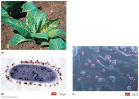
Viruses exhibit host specificity, infecting particular cell types within specific hosts. Some are generalists, infecting multiple cell types or species. All forms of life, including bacteria (bacteriophages), plants, animals, and fungi, are susceptible to viral infection.
Bacteriophages: Viruses that infect bacteria.
Plant viruses: Infect crops, often entering through wounds or via parasites.
Capsid Morphology
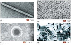
The capsid is composed of protein subunits called capsomeres, which protect the viral genome and facilitate attachment to host cells. Capsids can have various shapes, including helical, polyhedral, and complex forms.
Helical: Rod-shaped, as in tobacco mosaic virus.
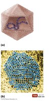
Polyhedral: Many-sided, as in adenovirus.
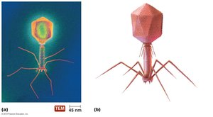
Complex: Bacteriophage T4 has a complex structure with a head, tail, and tail fibers.
The Viral Envelope
Some viruses acquire an envelope from the host cell membrane during replication or release. The envelope consists of a phospholipid bilayer and proteins, including virally encoded glycoproteins (spikes) that play a role in host recognition and attachment.
Enveloped viruses: More sensitive to environmental conditions than non-enveloped (naked) viruses.
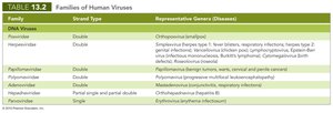
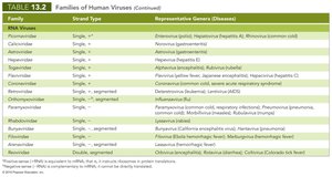
Classification of Viruses
Viruses are classified based on their nucleic acid type, presence or absence of an envelope, shape, and size. Unlike cellular organisms, viruses are not organized into kingdoms or phyla. The main taxonomic ranks are family (ending in -viridae), genus (ending in -virus), and species (often based on host).
Family | Strand Type | Representative Genera (Diseases) |
|---|---|---|
Herpesviridae | Double | Herpes simplex virus (cold sores, genital herpes) |
Orthomyxoviridae | Single, segmented | Influenza virus (influenza) |
Viral Replication
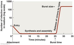
Lytic Replication Cycle
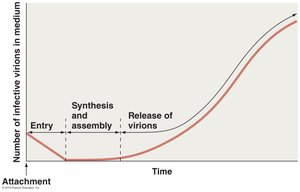
The lytic cycle is a process by which viruses replicate within a host cell, ultimately causing cell lysis and release of new virions. The five stages are:
Attachment: Virus binds to specific receptors on the host cell surface.
Entry: Viral genome enters the host cell.
Synthesis: Host machinery synthesizes viral components.
Assembly: New virions are assembled from synthesized components.
Release: Host cell lyses, releasing new virions.
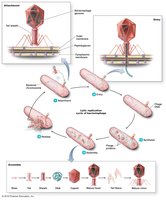
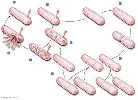
Lysogeny
Lysogeny is a modified replication cycle in which the viral genome integrates into the host chromosome as a prophage. The host cell survives and divides, passing the prophage to daughter cells. Induction can trigger the lytic cycle, leading to cell lysis.
Lysogenic conversion: Prophages may carry genes that alter the host phenotype (e.g., toxin production).
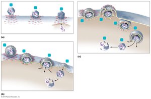
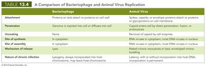
Replication of Animal Viruses
Animal viruses attach to host cells via glycoprotein spikes or other molecules. Entry and uncoating can occur by direct penetration, membrane fusion, or endocytosis. The replication strategy depends on the type of viral genome (DNA or RNA).
Direct penetration: Viral genome enters the cell directly (e.g., poliovirus).
Membrane fusion: Viral envelope fuses with host membrane (e.g., measles virus).
Endocytosis: Entire virion is engulfed by the host cell (e.g., herpesvirus).
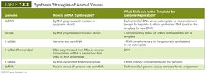
Synthesis of Animal Viruses
The synthesis of viral components varies by genome type:
dsDNA viruses: Replicate in the nucleus; proteins synthesized in the cytoplasm.
ssDNA viruses: Host enzymes synthesize a complementary DNA strand to form dsDNA.
RNA viruses: Replicate in the cytoplasm; strategies differ for +ssRNA, -ssRNA, and dsRNA viruses.
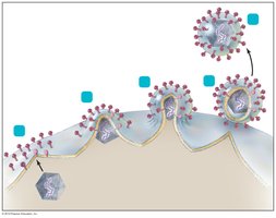
Assembly and Release of Animal Viruses
Most DNA viruses assemble in the nucleus, while RNA viruses assemble in the cytoplasm. Enveloped viruses are released by budding, which may result in persistent infections. Naked viruses are released by exocytosis or cell lysis.
Latency of Animal Viruses
Some animal viruses can remain dormant within host cells as latent viruses or proviruses. Latency may last for years without symptoms, and integration into host DNA is permanent.
Other Parasitic Particles: Viroids and Prions
Viroids
Viroids are extremely small, circular RNA molecules that infect plants. They lack a protein capsid and are pathogenic in many crops. Their origin is hypothesized to be related to introns.
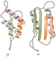
Prions
Prions are infectious proteins that cause neurodegenerative diseases. Normal cellular prion protein (PrP) has an α-helical structure, while the disease-causing form (prion PrP) has a β-sheet structure. Prion PrP induces normal PrP to refold into the pathogenic form, leading to spongiform encephalopathies.
Diseases: Bovine spongiform encephalopathy (BSE), variant Creutzfeldt-Jakob disease (vCJD), kuru.
Transmission: Ingestion, transplantation, or contact with infected tissues.
Destruction: Prions are destroyed by incineration or autoclaving in concentrated sodium hydroxide.
Culturing Viruses in the Laboratory
Methods of Culturing Viruses
Viruses cannot grow in standard microbiological media and must be cultured within host cells. Common methods include:
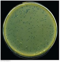
Cell culture: Viruses are grown in cultured cells, forming plaques where cells are lysed.
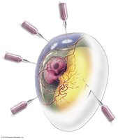
Embryonated eggs: Fertilized chicken eggs provide a sterile, nutrient-rich environment for viral growth; used in vaccine production.
Plants and animals: Used for viruses that do not grow well in cell culture or eggs.