Back
BackCharacterizing and Classifying Viruses, Viroids, and Prions
Study Guide - Smart Notes
 Tailored notes based on your materials, expanded with key definitions, examples, and context.
Characteristics of Viruses
Nature and Structure of Viruses
Viruses are minuscule, acellular infectious agents that contain either DNA or RNA as their genetic material, but never both. They are not considered living organisms because they cannot carry out metabolic processes, do not grow or respond to the environment, and cannot reproduce independently. Viruses lack a cytoplasmic membrane, cytosol, and organelles.
Obligate intracellular parasites: Viruses must infect a host cell to replicate, utilizing the host's organelles and enzymes.
Diseases: Viruses are responsible for many human diseases, including influenza, HIV, rabies, hepatitis, and emerging diseases like Zika and Ebola.
Role in cancer: It is estimated that viruses cause about 25% of human cancers.
Viral States: Extracellular and Intracellular
Viruses exist in two states:
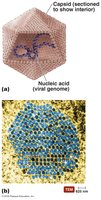
Extracellular state (virion): The complete virus particle, consisting of a nucleic acid genome surrounded by a protein coat (capsid), and sometimes a phospholipid envelope. The nucleic acid and capsid together are called the nucleocapsid. The outermost layer provides protection and recognition sites for host cells.
Intracellular state: The capsid is removed, and the virus exists as nucleic acid within the host cell.
Host Range and Specificity
Most viruses infect only specific hosts or even specific cell types within a host, due to the affinity between viral surface proteins and host cell receptors. Some viruses are highly specific (e.g., HIV infects only human helper T cells), while others are generalists (e.g., West Nile virus infects multiple species).
Bacteriophages: Viruses that infect bacteria.
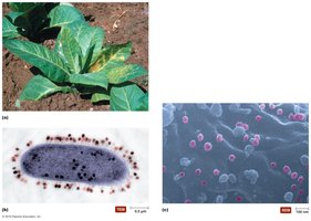
Plant viruses: Infect many food crops.
Fungal viruses: Less studied, often lack an extracellular state.
Genetic Material of Viruses
Viral genomes are much smaller than those of cells and show greater variety. They may be DNA or RNA, single- or double-stranded, linear or circular, and segmented or non-segmented. The type of nucleic acid is a primary criterion for virus classification. 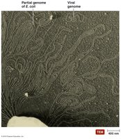
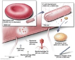
Size and Morphology
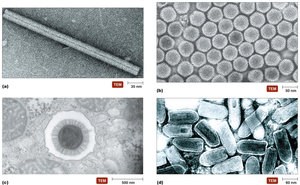
Most viruses range from 20 to 400 nm in diameter. Only the largest viruses can be seen with a light microscope. Viral capsids, made of protein subunits called capsomeres, protect the genome and facilitate attachment to host cells. Capsid shapes include:
Helical: Capsomeres arranged in a spiral (e.g., tobacco mosaic virus).
Polyhedral: Spherical, often icosahedral (20-sided).
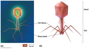
Complex: Structures that do not fit into the other categories (e.g., bacteriophage T4).
Viral Envelope
Some animal viruses have an envelope derived from the host cell membrane, composed of a phospholipid bilayer and proteins (often with glycoprotein spikes). The envelope aids in host recognition and entry. Viruses lacking an envelope are called naked virions. 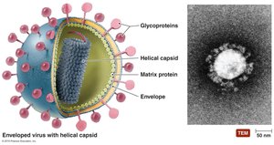
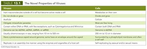
Comparison of Viruses and Cells
Viruses | Cells |
|---|---|
Obligate intracellular parasites | Free-living |
Contain either DNA or RNA | Contain both DNA and RNA |
Acellular | Cellular |
20–400 nm in size | 200 nm to several meters in diameter |
Replicate by assembly using host enzymes | Self-replicating by binary fission or mitosis |
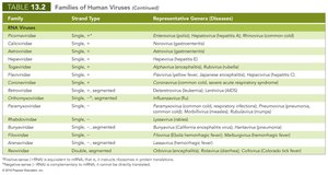
Classification of Viruses
Taxonomy and Criteria
The International Committee on Taxonomy of Viruses classifies viruses based on nucleic acid type, presence of envelope, shape, and size. Unlike cellular organisms, viruses are not classified into kingdoms or divisions. 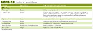
Viral Replication
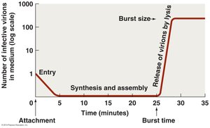
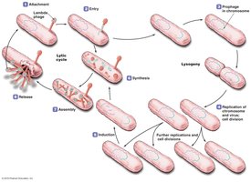
Lytic Replication Cycle
Viruses depend on host cell machinery for replication. The lytic cycle, typical of many bacteriophages, results in host cell lysis and release of new virions. The five stages are:
Attachment: Virus attaches to host cell via specific interactions.
Entry: Viral genome enters the host, often with the help of enzymes like lysozyme.
Synthesis: Host machinery is redirected to synthesize viral components.
Assembly: New virions are assembled from synthesized components.
Release: Host cell lyses, releasing new virions.
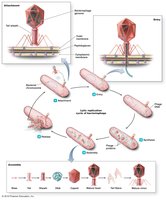
Lysogeny (Temperate Phages)
Some bacteriophages can integrate their genome into the host chromosome, becoming a prophage. The host cell survives and divides, passing the prophage to daughter cells. Environmental triggers can induce the prophage to enter the lytic cycle.
Lysogenic conversion: Prophages may carry genes that alter the host phenotype, sometimes turning harmless bacteria into pathogens.
Replication of Animal Viruses
Animal viruses follow similar steps as bacteriophages but differ due to the presence of envelopes, the eukaryotic nature of host cells, and the lack of a cell wall. Entry mechanisms include:
Direct penetration (e.g., poliovirus)
Membrane fusion (e.g., measles virus)
Endocytosis (e.g., herpes simplex virus)
Capsids are removed (uncoating) before replication. 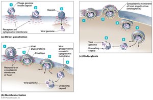
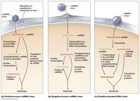
Synthesis Strategies in Animal Viruses
Replication strategies depend on the type of viral genome:
dsDNA viruses: Replicate in the nucleus; proteins made in cytoplasm.
ssDNA viruses: Host enzymes synthesize a complementary DNA strand to form dsDNA.
+ssRNA viruses: Genome acts as mRNA and is directly translated.
-ssRNA viruses: Require RNA-dependent RNA transcriptase to synthesize +RNA.
dsRNA viruses: Positive strand is translated; negative strand serves as template.
Retroviruses: +ssRNA is reverse transcribed into DNA (e.g., HIV).
Assembly and Release of Animal Viruses
Most DNA viruses assemble in the nucleus, while RNA viruses assemble in the cytoplasm. Enveloped viruses are released by budding, allowing persistent infections. Naked viruses are released by exocytosis or lysis. 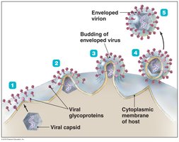
Latency in Animal Viruses
Some animal viruses can remain dormant (latent) within host cells for years. Latent viruses (proviruses) may integrate into the host genome, and this incorporation is permanent for some viruses.
The Role of Viruses in Cancer
Oncogenes and Tumor Formation
Cell division is tightly regulated by genes. Neoplasia (uncontrolled cell division) leads to tumor formation. Viruses can contribute to cancer by:
Carrying oncogenes
Promoting host oncogenes
Interfering with tumor suppressor genes
Examples of virus-induced cancers include Burkitt’s lymphoma, Hodgkin’s disease, Kaposi’s sarcoma, and cervical cancer. 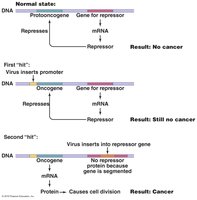
Culturing Viruses in the Laboratory
Methods of Culturing Viruses
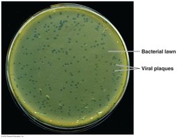
Viruses require living cells for growth and are cultured in:
Mature organisms: Bacteriophages are grown in bacteria, producing plaques (clear zones of lysed cells).
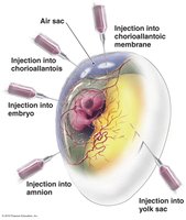
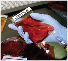
Embryonated eggs: Fertilized chicken eggs provide a sterile, nutrient-rich environment for viral growth and vaccine production.
Cell cultures: Cells isolated from tissues are grown in vitro. Diploid cell cultures are short-lived; continuous cell cultures (from cancer cells) can divide indefinitely.
Viroids and Prions
Viroids
Viroids are extremely small, circular RNA molecules that infect plants. They lack a protein capsid and are similar to RNA viruses. Viroid-like agents can also infect fungi. 
Prions
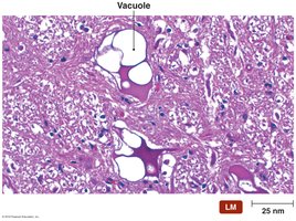
Prions are infectious proteins that lack nucleic acids. They cause normal cellular proteins (PrP) to misfold, forming aggregates that damage neural tissue, resulting in spongiform encephalopathies (e.g., mad cow disease, vCJD, kuru, chronic wasting disease). Prions are highly resistant to standard decontamination methods. 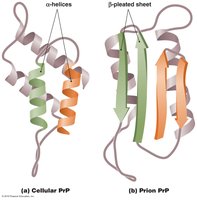
Comparison of Viruses, Viroids, and Prions
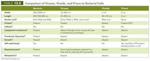
Bacteria | Viruses | Viroids | Prions | |
|---|---|---|---|---|
Width | 200–2000 nm | 10–400 nm | 2–3 nm | 5 nm |
Length | 300–10,000 nm | 20–1000 nm | 246–375 nucleotides | — |
Nucleic acid? | Both DNA and RNA | Either DNA or RNA, never both | RNA only | None |
Capsid? | Present | Present | Absent | Absent |
Growth? | Yes | No | No | No |
Self-replicating? | Yes | Yes, but only in host cell | No | Yes, transform PrP protein already present in cell |
Metabolism? | Present | Absent | Absent | Absent |