 Back
BackCharacterizing and Classifying Viruses, Viroids, and Prions: Study Notes
Study Guide - Smart Notes
 Tailored notes based on your materials, expanded with key definitions, examples, and context.
Characterizing and Classifying Viruses, Viroids, and Prions
Characteristics of Viruses
Viruses are minuscule, acellular infectious agents that possess either DNA or RNA as their genetic material. They are responsible for numerous diseases affecting humans, animals, plants, and bacteria. Unlike cellular organisms, viruses cannot carry out metabolic pathways, grow, or respond to their environment independently. Instead, they rely on host cells to reproduce and increase their numbers. Viruses lack cytoplasmic membranes, cytosol, and organelles, and exist in two states: extracellular (virion) and intracellular (nucleic acid only).
Virion: The extracellular state, consisting of nucleic acid surrounded by a protein coat (capsid), and sometimes a phospholipid envelope.
Intracellular State: The virus exists as nucleic acid after the envelope and capsid are removed.
Host Specificity: Most viruses infect only particular host cells due to the affinity of viral surface proteins for complementary proteins on the host cell surface. Some viruses are generalists, infecting many kinds of cells or hosts.
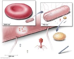
Figure: Relative sizes of cells, viruses, and other biological structures.
Are Viruses Alive?
There is ongoing debate among scientists regarding the status of viruses as living entities. Viruses exhibit some characteristics of life, such as possessing their own genomes, evolving over time, and invading host cells to take control. However, they lack independent growth, self-reproduction, responsiveness, and metabolism.
Living Characteristics: Ability to invade cells, control host cells, possess genomes, and evolve.
Non-living Characteristics: Cannot grow, reproduce, or metabolize independently.
Novel Properties of Viruses vs. Cells
Viruses differ fundamentally from cells in several ways, including their structure, genetic material, and replication strategies.
Viruses | Cells |
|---|---|
Inert outside of host cell; become active inside a cell | Metabolize on their own |
Do not divide or grow | Divide and grow |
Acellular | Cellular |
Obligate intracellular parasites | Can be free-living or parasitic |
Contain either DNA or RNA | Contain both DNA and RNA |
Usually chromosome is one piece, ranging from 10 nm to over 500 nm | 200 nm to 12 cm in diameter |
Have a proteinaceous capsid around genome, some have an envelope | Surrounded by a phospholipid membrane and often a cell wall |
Replicate in an assembly-line manner using host enzymes and organelles | Self-replicating by asexual and/or sexual means |
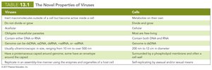
Table: Comparison of the novel properties of viruses and cells.
Genetic Material of Viruses
Viruses contain either DNA or RNA, which may be double-stranded (ds) or single-stranded (ss), and can be linear or circular. The type of nucleic acid is a primary criterion for virus classification. Viral genomes are much smaller than those of cells and contain very few genes.
Capsid Morphology and Shape
The capsid is a protein coat composed of subunits called capsomeres. Capsid shape is used in virus classification and can be helical, polyhedral, or icosahedral (20 sides). The capsid protects viral nucleic acid and facilitates attachment to host cells.
Viral Envelope
Some viruses acquire an envelope from the host cell during replication or release. The envelope consists of a phospholipid bilayer and proteins, including virally coded glycoproteins (spikes) that play a role in host recognition. Enveloped viruses are more fragile than naked viruses, but the envelope provides some protection from the immune system.
Classification of Viruses
Viruses are classified based on their nucleic acid type, presence of an envelope, shape, and size. Viral genera are organized into families, but relationships among viruses are not fully understood.
Families of Human Viruses
Human viruses are categorized into DNA and RNA viruses, each with distinct families and representative genera or diseases.
Family | Strand Type | Representative Genera (Disease) |
|---|---|---|
Parvoviridae | Double | Orthopoxvirus (smallpox) |
Herpesviridae | Double | Simplexvirus (herpes type 1 & 2), Varicellovirus (chickenpox), Epstein-Barr virus (infectious mononucleosis, Burkitt's lymphoma), Cytomegalovirus (birth defects), Roseolovirus (roseola) |
Papillomaviridae | Double | Papillomavirus (warts, cervical and penile cancers) |
Adenoviridae | Double | Mastadenovirus (pneumonia, respiratory infections) |
Hepadnaviridae | Double and single | Orthohepadnavirus (hepatitis B) |
Polyomaviridae | Double | Polyomavirus (multifocal leukoencephalopathy) |
Reoviridae | Double | Cytorhabdovirus (rotavirus) |
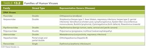
Table: Families of human DNA viruses.
Family | Strand Type | Representative Genera (Disease) |
|---|---|---|
Picornaviridae | Single, + | Enterovirus (polio), Hepatovirus (hepatitis A) |
Caliciviridae | Single, + | Norovirus (gastroenteritis) |
Hepeviridae | Single, + | Hepevirus (hepatitis E) |
Astroviridae | Single, + | Astrovirus (gastroenteritis) |
Coronaviridae | Single, + | Coronavirus (common cold, SARS, MERS) |
Flaviviridae | Single, + | Flavivirus (yellow fever, dengue, hepatitis C) |
Retroviridae | Single, + | Lentivirus (HIV) |
Paramyxoviridae | Single, - | Paramyxovirus (measles, mumps, respiratory infections) |
Rhabdoviridae | Single, - | Lyssavirus (rabies) |
Filoviridae | Single, - | Ebolavirus (Ebola) |
Orthomyxoviridae | Single, - segmented | Influenzavirus (influenza) |
Bunyaviridae | Single, - segmented | Bunyavirus (California encephalitis, hantavirus) |
Arenaviridae | Single, - segmented | Arenavirus (hemorrhagic fever, Lassa fever) |
Reoviridae | Double, segmented | Rotavirus (gastroenteritis) |
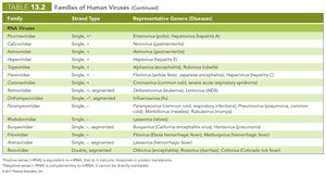
Table: Families of human RNA viruses.
Viral Attachment and Replication Cycle
Viruses depend on host organelles and enzymes to produce new virions. The lytic replication cycle results in the death and lysis of the host cell and consists of five stages: attachment, entry, synthesis, assembly, and release.
Attachment: Virus binds to host cell surface.
Entry: Viral genome enters host cell.
Synthesis: Host cell machinery produces viral components.
Assembly: New virions are assembled.
Release: Virions are released, often causing cell lysis.
Lysogenic Replication Cycle (Lysogeny)
Some bacteriophages undergo a modified replication cycle called lysogeny, where infected host cells grow and reproduce normally for generations before lysing. Temperate phages integrate their genome into the host chromosome as a prophage, which can later be induced to enter the lytic cycle. Lysogenic conversion occurs when phages carry genes that alter the phenotype of a bacterium.
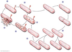
Figure: The lysogenic replication cycle in bacteriophages.
Replication of Animal Viruses
Animal viruses follow a similar basic replication pathway as bacteriophages, but differences arise due to the presence of an envelope, the eukaryotic nature of animal cells, and the lack of a cell wall. Attachment is mediated by glycoprotein spikes or other molecules, and entry can occur via direct penetration, membrane fusion, or endocytosis.
Direct Penetration: Viral genome enters directly.
Membrane Fusion: Enveloped virus fuses with host membrane.
Endocytosis: Host cell engulfs virus.
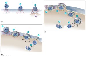
Figure: Three mechanisms of entry of animal viruses.
Synthesis of DNA and RNA Viruses in Animals
Animal viruses use different strategies for synthesis depending on their nucleic acid type. DNA viruses often enter the nucleus, while RNA viruses replicate in the cytoplasm. Positive-sense (+) RNA can act as mRNA, while negative-sense (–) RNA must be transcribed before translation. Retroviruses use reverse transcription to synthesize DNA from RNA.
dsDNA Viruses: Replicate genome in nucleus; proteins made in cytoplasm.
ssDNA Viruses: ssDNA folds to form dsDNA, which is replicated.
+ssRNA Viruses: RNA acts as mRNA.
–ssRNA Viruses: RNA must be transcribed to +ssRNA.
Retroviruses: Use reverse transcriptase to synthesize DNA from RNA.
Assembly and Release of Animal Viruses
Most DNA viruses assemble in the nucleus, while most RNA viruses assemble in the cytoplasm. The number of viruses produced depends on the virus type and host cell health. Enveloped viruses cause persistent infections, while naked viruses are released by exocytosis or lysis.
Latency of Animal Viruses
Some animal viruses remain dormant in host cells as latent viruses or proviruses. Latency can be prolonged for years with no viral activity, and incorporation of provirus into host DNA is permanent. Examples include HIV, chickenpox, hepatitis B, and herpes virus.
Role of Viruses in Cancer
Viruses can contribute to cancer by disrupting genetic control of cell division. Neoplasia is uncontrolled cell division, resulting in tumors. Malignant tumors (cancers) can metastasize. Viruses cause 20–25% of human cancers, either by carrying oncogenes, promoting host oncogenes, or interfering with tumor repression. Examples include Burkitt's lymphoma, Hodgkin's disease, Kaposi's sarcoma, and cervical cancer.
Culturing Viruses in the Laboratory
Viruses cannot grow in standard microbiological media and must be cultured inside host cells. Three types of media are used: mature organisms (bacteria, plants, animals), embryonated eggs, and cell cultures.
Viroids
Characteristics of Viroids
Viroids are extremely small, circular pieces of single-stranded RNA that are infectious and pathogenic in plants. Unlike viruses, viroids lack a capsid and do not code for proteins. Viroid RNA adheres to complementary plant RNA, leading to degradation by plant enzymes and resulting in disease.
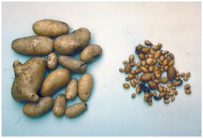
Figure: Potato plants affected by viroids.
Prions
Characteristics of Prions
Prions are proteinaceous infectious agents found in mammals. Normal cellular prion protein (PrP) has α-helices, while disease-causing prion PrP has β-sheets. Prion PrP induces refolding of cellular PrP into the disease-causing form.
Prion Diseases
Prion diseases, known as spongiform encephalopathies, are characterized by large vacuoles in brain tissue, giving it a spongy appearance. Examples include bovine spongiform encephalopathy (BSE), scrapie, kuru, chronic wasting disease (CWD), and variant Creutzfeldt-Jakob disease (vCJD). Prion diseases are transmitted by ingestion, transplantation, or contact with infected tissues, and there is no standard treatment.
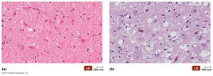
Figure: Brain tissue affected by prion disease.
Prion Sterilization
Normal sterilization procedures do not deactivate prions. Prions are destroyed by incineration or autoclaving in concentrated sodium hydroxide. The European Union has approved the use of enzymes to remove prions from medical equipment.
