 Back
BackClassification and Identification of Microorganisms & Microscopy in Microbiology
Study Guide - Smart Notes
 Tailored notes based on your materials, expanded with key definitions, examples, and context.
Classification and Identification of Microorganisms
Overview of Microbial Classification
The classification of microorganisms is fundamental to understanding their diversity, relationships, and roles in nature and disease. Microorganisms are grouped based on shared characteristics, genetic relationships, and ecological roles.
Prokaryotic species: Populations of cells with similar characteristics. Subdivisions include:
Culture: Bacteria grown in laboratory media.
Clone: Population of cells derived from a single parent cell.
Strain: Genetically different cells within a clone.
Protista: A diverse kingdom including autotrophic and heterotrophic organisms, grouped into clades based on rRNA.
Fungi: Chemoheterotrophic, unicellular or multicellular, with chitin cell walls; reproduce via spores or hyphal fragments.
Plantae: Multicellular, cellulose cell walls, photosynthetic.
Animalia: Multicellular, no cell walls, chemoheterotrophic.
Viral species: Populations of viruses with similar characteristics occupying a particular ecological niche; not classified within domains as they are not cellular and require host cells for replication.
Classification vs. Identification
Classification and identification are distinct but related processes in microbiology.
Classification: Placing organisms into groups of related species based on lists of characteristics.
Identification: Matching characteristics of an unknown organism to known organisms, often for clinical or research purposes.
Methods of Classifying and Identifying Microorganisms
Microorganisms can be classified and identified using a variety of morphological, biochemical, and molecular techniques.
Morphological characteristics: Useful for identifying eukaryotes but provide limited phylogenetic information.
Differential staining: Includes Gram staining and acid-fast staining; not useful for bacteria lacking cell walls.
Biochemical tests: Determine the presence of specific bacterial enzymes and metabolic pathways.
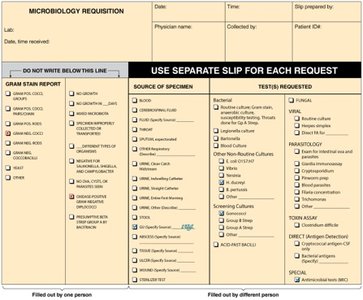
Example: A clinical microbiology lab requisition form is used to request and document tests for identifying pathogens in patient samples.
Biochemical Identification of Bacteria
Biochemical tests are essential for distinguishing among bacterial genera and species, especially among enteric bacteria.
Tests include fermentation of sugars, utilization of specific carbon sources, and production of metabolic byproducts.
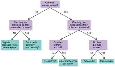
Example: The ability to ferment lactose, use citric acid, or produce acetoin helps differentiate Escherichia coli, Citrobacter, and Enterobacter.
Rapid Identification Methods
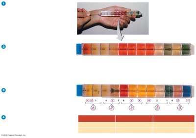
Modern clinical labs use rapid identification systems that combine multiple biochemical tests in a single device.
Example: The EnteroPluri Test contains media for 15 biochemical tests. After inoculation and incubation, results are scored and compared to a database for identification.
Serological Methods
Serological tests use antibodies to detect specific microbial antigens, allowing for differentiation at the species or strain level.
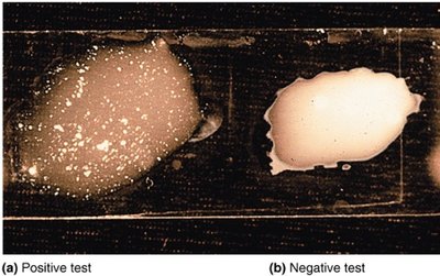
Slide agglutination test: Bacteria agglutinate (clump) when mixed with specific antibodies, indicating a positive reaction.
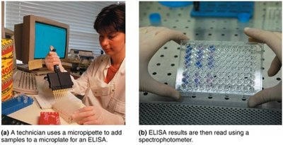
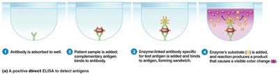
Enzyme-Linked Immunosorbent Assay (ELISA)
ELISA is a sensitive method for detecting microbial antigens or antibodies in clinical samples.
Known antibodies are used to capture and detect unknown bacteria or their products.
Results are visualized by a color change, which can be measured spectrophotometrically.
Microscopy in Microbiology
Units of Measurement
Microorganisms are measured in micrometers (μm) and nanometers (nm).
1 μm = 10-6 m
1 nm = 10-9 m
Types of Light Microscopy
Light microscopy uses visible light to observe specimens. Several types are used in microbiology:
Compound light microscopy
Darkfield microscopy
Phase-contrast microscopy
Differential interference contrast (DIC) microscopy
Fluorescence microscopy
Confocal microscopy
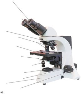
Compound Light Microscope: Structure and Function
The compound light microscope is the most commonly used microscope in microbiology. It uses a series of lenses to magnify specimens.
Ocular lens (eyepiece): Remagnifies the image formed by the objective lens.
Objective lenses: Primary lenses that magnify the specimen.
Stage: Holds the microscope slide in position.
Condenser: Focuses light through the specimen.
Diaphragm: Controls the amount of light entering the condenser.
Illuminator: Light source.
Coarse and fine focusing knobs: Adjust the focus of the image.
Magnification and Resolution
Magnification is the product of the objective and ocular lens powers. Resolution is the ability to distinguish two points as separate.
Total magnification:
Resolution: A microscope with a resolving power of 0.4 nm can distinguish between two points at least 0.4 nm apart.
Shorter wavelengths of light provide greater resolution.
Oil Immersion Technique
Immersion oil is used with high-power objective lenses to reduce light refraction and increase resolution.
Refractive index: A measure of the light-bending ability of a medium.
Immersion oil prevents loss of light due to refraction, allowing more light to enter the objective lens.
Preparation and Staining of Specimens
Staining enhances contrast in microscopic specimens, making cellular structures more visible.
Smear: A thin film of microorganisms spread on a slide and fixed by heat or chemicals.
Stains: Consist of a colored ion (chromophore). Basic dyes have a positively charged chromophore; acidic dyes have a negatively charged chromophore.
Negative staining: Stains the background, not the cell.
Simple stain: Uses a single basic dye to highlight the entire microorganism.
Mordant: Used to hold the stain or enlarge the specimen.
Example: Gram staining differentiates bacteria into Gram-positive and Gram-negative based on cell wall structure.
Key Equations
Total Magnification:
Summary Table: Methods for Identifying Microorganisms
Method | Principle | Application |
|---|---|---|
Morphological characteristics | Observation of cell shape, size, and arrangement | Useful for eukaryotes, limited for bacteria |
Differential staining | Gram, acid-fast stains | Distinguishes major groups of bacteria |
Biochemical tests | Detection of metabolic enzymes | Identification of bacterial species |
Serological methods | Antibody-antigen reactions | Species and strain identification |
Molecular methods | DNA sequencing, PCR, hybridization | Precise identification and classification |
Additional info: Molecular methods such as PCR and DNA sequencing are increasingly important for identifying microbes that are difficult to culture or distinguish by traditional methods.
