 Back
BackColony Morphology: Describing Microbial Colonies on Agar Plates
Study Guide - Smart Notes
 Tailored notes based on your materials, expanded with key definitions, examples, and context.
Colony Morphology in Microbiology
Introduction
Colony morphology refers to the observable characteristics of microbial colonies grown on solid media, such as agar plates. Accurate description of these features is essential for identification and classification of microorganisms in microbiology. Standardized terminology ensures clear communication among scientists and students.
Culture Conditions
The appearance of microbial colonies can be influenced by the conditions under which they are cultured. It is important to document:
Medium: Specify the type of agar used (e.g., R2A agar, Tryptic soy agar).
Temperature: Record the incubation temperature.
Age: Note the age of the culture at the time of observation.
These factors can affect colony size, color, and other morphological features.
Colony Form
Colony form describes the overall shape and structure of the colony. Common forms include:
Circular: Round colonies with smooth edges.
Irregular: Colonies with uneven, non-circular shapes; may spread rapidly.
Filamentous: Colonies with thread-like extensions.
Rhizoid: Colonies with branching, root-like structures.
Punctiform: Very small, dot-like colonies.
Spindle: Lens-shaped colonies.
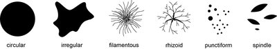
Example: Bacillus subtilis often forms irregular, spreading colonies, while Staphylococcus aureus forms circular colonies.
Colony Elevation
Elevation describes the vertical profile of the colony. Common types include:
Flat (circular): Colony lies flat against the agar.
Raised: Slightly elevated above the agar surface.
Convex: Dome-shaped, semi-circular in cross-section.
Pulvinate: Deeply convex, higher than convex.
Umbonate: Raised with a central bump (umbo).
Crateriform: Colony with a central depression.
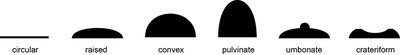
Example: Micrococcus luteus often forms convex colonies, while Pseudomonas aeruginosa may form flat colonies.
Colony Margin
Margin refers to the edge of the colony. Types include:
Entire: Smooth, even edge.
Undulate: Wavy edge.
Filamentous: Edge with thread-like extensions.
Lobate: Edge with deep indentations.
Erose (serrated): Jagged, irregular edge.
Curled: Edge with concentric rings.
Scalloped: Edge with rounded projections.
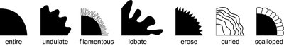
Example: Streptomyces species often have filamentous margins.
Colony Surface
The surface of a colony can be described as:
Smooth: Even, glossy surface.
Glistening: Shiny appearance.
Rough: Uneven, coarse surface.
Wrinkled: Surface with folds or ridges.
Dry: Lacks moisture.
Powdery: Appears dusted.
Moist: Appears wet.
Mucoid: Large, moist, sticky colonies.
Brittle: Breaks easily.
Viscous: Difficult to remove from loop.
Butyrous: Buttery texture.
Example: Klebsiella pneumoniae forms mucoid colonies due to capsule production.
Colony Opacity
Opacity describes how much light passes through the colony:
Transparent: Clear, allows light to pass through.
Translucent: Partially allows light through.
Opaque: Blocks transmitted light.
Check opacity using a dissecting microscope for accuracy.
Colony Color
Color is an important distinguishing feature. Colonies may be:
White
Buff
Brown
Red
Yellow
Pink
Purple
Other colors
Example: Serratia marcescens produces red colonies due to pigment production.
Other Distinguishing Characteristics
Additional features to note include:
Odor: Some colonies produce distinctive smells.
Diffusible pigments: Pigments that stain the agar.
Other characteristics: Any unique features not covered above.
Example: Pseudomonas aeruginosa produces a fruity odor and blue-green pigment that diffuses into the agar.
