 Back
BackComprehensive Study Notes: Classification, Pathogenicity, and Immunity in Microbiology
Study Guide - Smart Notes
 Tailored notes based on your materials, expanded with key definitions, examples, and context.
Classification of Prokaryotes: Bacteria and Archaea
Gram-Negative Bacteria
Gram-negative bacteria are a diverse group of prokaryotes with a thin peptidoglycan layer and an outer membrane containing lipopolysaccharides. They are classified into several major groups, many of which are clinically significant.
Betaproteobacteria: Includes Neisseria species, such as N. gonorrhoeae (gonorrhea) and N. meningitidis (meningitis).
Gammaproteobacteria: Contains important pathogens like Escherichia coli, Pseudomonas aeruginosa, and Klebsiella species, associated with urinary tract infections, pneumonia, and healthcare-associated infections.
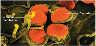
Chlamydiae: Includes Chlamydia trachomatis, which causes sexually transmitted infections, pneumonia, and eye infections.
Bacteroidetes: Part of the normal human microbiota but can cause infections in immunocompromised patients, especially in abdominal and pelvic regions.
Spirochaetia: Includes Treponema pallidum (syphilis), Borrelia species (Lyme disease), and Leptospira species.
Additional info: Other classes such as Alphaproteobacteria, Deltaproteobacteria, Campylobacterales, Cyanobacteria, Chlorobia, Chloroflexi, Planctomycetia, and Fusobacteria are less commonly encountered in clinical practice but may be relevant in specific infections.
Pseudomonadota
Pseudomonadota is the largest taxonomic group of bacteria, mostly chemoheterotrophic, and capable of surviving in low-nutrient environments. Many have stalks or buds called prosthecae.
Spirillum: Found in freshwater, moves via polar flagella.
Sphaerotilus: Found in freshwater and sewage, forms protective sheaths.
Selected Pathogenic Genera
Burkholderia: B. cepacia degrades organic molecules; B. pseudomallei causes melioidosis.
Bordetella: B. pertussis causes whooping cough.
Neisseria: N. gonorrhoeae (gonorrhea), N. meningitidis (meningitis).
Zoogloea: Important in wastewater treatment (activated sludge system).
Chlamydiota – Chlamydiae
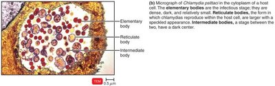
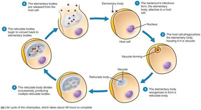
Chlamydiae are obligate intracellular bacteria lacking peptidoglycan in their cell walls. They have a unique developmental cycle involving infectious elementary bodies and replicative reticulate bodies.
Chlamydia trachomatis: Causes trachoma (eye infection) and urethritis.
Chlamydophila psittaci: Causes respiratory psittacosis (parrot fever).
Chlamydophila pneumoniae: Causes mild pneumonia in young adults.
Bacteroidota – Bacteroidetes
Bacteroidetes are anaerobic bacteria commonly found in the mouth and large intestine. Bacteroides fragilis is part of the normal flora but can cause infections if displaced.
Spirochaetota – Spirochaetia
Spirochetes are spiral-shaped bacteria that move via axial filaments. Notable genera include:
Treponema: T. pallidum causes syphilis.
Borrelia: Causes relapsing fever and Lyme disease (bull’s eye rash).
Leptospira: Excreted in animal urine, can infect humans.
Gram-Positive Bacteria
G+C Ratio and Classification
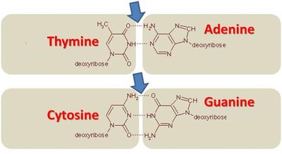
The G+C ratio is the percentage of guanine and cytosine bases in DNA. It is used to classify bacteria and infer evolutionary relationships.
High G+C: Streptomyces (69–73%), Mycobacterium (62–70%)
Low G+C: Streptococcus (33–44%), Clostridium (21–54%)
Bacillota – Bacilli
Bacillus: Endospore-forming rods; B. anthracis (anthrax), B. thuringiensis (biopesticide), B. cereus (food poisoning).
Staphylococcus: Grape-like clusters; S. aureus causes wound infections, is often antibiotic-resistant, and produces enterotoxins.
Lactobacillales
Lactobacillus: Aerotolerant anaerobes, produce lactic acid, important in food production and microbiota.
Streptococcus: Chains of cocci; S. pyogenes (strep throat), S. pneumoniae (pneumonia), S. mutans (dental caries).
Enterococcus: Found in the intestinal tract; E. faecalis and E. faecium cause hospital-acquired infections.
Listeria: L. monocytogenes contaminates food, causes listeriosis.
Clostridota – Clostridia
Clostridium: Endospore-forming obligate anaerobes; C. tetani (tetanus), C. botulinum (botulism), C. perfringens (gas gangrene).
Clostridioides difficile: Causes antibiotic-associated diarrhea and colitis.
Epulopiscium: Large bacteria, unique reproduction (daughter cells form within parent cell).
Mycoplasmatota
Mycoplasma: Lack cell wall, resistant to antibiotics targeting cell wall synthesis; M. pneumoniae causes mild pneumonia.
Actinomycetota
Mycobacterium: M. tuberculosis (tuberculosis).
Corynebacterium: C. diphtheriae (diphtheria).
Gardnerella: G. vaginalis (vaginitis).
Actinomyces: A. israelii (actinomycosis).
Deinococcota
Deinococcus radiodurans: Highly resistant to radiation, used in bioremediation.
Thermus aquaticus: Source of Taq polymerase for PCR.
Archaea
Archaea are prokaryotes lacking peptidoglycan, often extremophiles (halophiles, thermophiles, acidophiles). Methanogens produce methane and are part of the human microbiota.
The Eukaryotes: Fungi, Algae, Protozoa, and Helminths
Fungi
Fungi are eukaryotic, chemoheterotrophic organisms with cell walls containing chitin. They reproduce sexually and asexually via spores and play roles in decomposition, food production, and disease.
Vegetative Structures: Thallus (body), hyphae (filaments), mycelium (mass of hyphae).
Yeasts: Unicellular, reproduce by budding or fission.
Dimorphic Fungi: Exist as molds or yeasts depending on conditions.
Medically Important Fungi:
Mucoromycota: Mucor, Rhizopus (mucormycosis).
Ascomycota: Candida (candidiasis), Aspergillus (aspergillosis).
Basidiomycota: Cryptococcus neoformans (cryptococcosis).
Lichens
Lichens are mutualistic associations between a fungus and an alga or cyanobacterium. They are important in ecosystems and have economic uses (dyes, litmus, antimicrobial compounds).
Algae
Algae are photoautotrophic, aquatic organisms lacking true roots, stems, and leaves. They reproduce sexually and asexually and are important in aquatic food webs and oxygen production.
Protozoa
Protozoa are unicellular eukaryotes, often motile, and can be free-living or parasitic. They reproduce asexually (fission, budding, schizogony) and sometimes sexually.
Excavata: Giardia intestinalis, Trichomonas vaginalis, Trypanosoma (sleeping sickness).
Amoebozoa: Entamoeba histolytica (amebic dysentery), Acanthamoeba, Balamuthia.
Apicomplexa: Toxoplasma gondii, Cryptosporidium, Plasmodium (malaria).
Ciliates: Balantidium coli (dysentery).
Helminths
Helminths are multicellular parasitic worms, including flatworms (Platyhelminthes) and roundworms (Nematoda). They have complex life cycles and can cause significant human disease.
Arthropods as Vectors
Arthropods (insects, arachnids) can transmit pathogens mechanically or biologically. Examples include mosquitoes (malaria), ticks (Lyme disease), and fleas (plague).
Microbial Mechanisms of Pathogenicity
Pathogenicity and Virulence
Pathogenicity is the ability to cause disease; virulence is the degree of pathogenicity. Pathogens enter hosts via mucous membranes, skin, or parenteral routes. The infectious dose (ID50) and lethal dose (LD50) quantify virulence and toxin potency, respectively.
Adherence and Invasion
Pathogens attach to host cells using adhesins (e.g., glycocalyx, fimbriae, viral spikes). Capsules and cell wall components (M protein, Opa protein, mycolic acid) help evade immune defenses. Enzymes (coagulases, kinases, hyaluronidase, collagenase, IgA proteases) facilitate invasion and immune evasion.
Antigenic Variation
Some pathogens alter their surface antigens to evade immune detection (e.g., influenza virus, Neisseria gonorrhoeae, Trypanosoma brucei).
Biofilms
Biofilms protect bacteria from antibiotics and immune cells, contributing to chronic infections.
Siderophores
Pathogens secrete siderophores to acquire iron from the host, essential for bacterial growth.
Direct Damage and Toxins
Pathogens can damage host cells directly or via toxins. Exotoxins are secreted proteins (often by Gram-positive bacteria), while endotoxins are lipopolysaccharides (LPS) from Gram-negative bacteria released upon cell lysis.
Feature | Exotoxins | Endotoxins |
|---|---|---|
Source | Secreted by living bacteria (mainly Gram+) | Part of Gram- cell wall; released on lysis |
Chemical Nature | Proteins/peptides | Lipopolysaccharides (LPS) |
Heat Stability | Heat-sensitive | Heat-stable |
Toxicity | Highly toxic | Less toxic |
Immunogenicity | Highly immunogenic | Poorly immunogenic |
Specificity | Often tissue-specific | Systemic effects |
Examples | Botulinum, tetanus toxins | LPS from Gram- bacteria |
Types of Exotoxins
A-B Toxins: Two-part toxins (A = active, B = binding). Example: Diphtheria toxin inhibits protein synthesis.
Membrane-Disrupting Toxins: Form pores in host membranes (e.g., hemolysins, leukocidins).
Superantigens: Overstimulate T cells, causing cytokine storm (e.g., toxic shock syndrome toxin).
Endotoxins
Lipid A of LPS triggers strong immune responses, fever, and can cause endotoxic shock. Detected by the Limulus Amebocyte Lysate (LAL) assay.
Plasmids and Lysogeny
Plasmids can carry virulence genes (antibiotic resistance, toxin production). Lysogenic conversion by bacteriophages can alter bacterial properties.
Food Infection vs. Food Intoxication
Aspect | Food Infection | Food Intoxication |
|---|---|---|
Cause | Ingesting live bacteria | Ingesting pre-formed toxins |
Toxin Production | In body after infection | In food before ingestion |
Onset | 6–72 hours | Within hours |
Examples | Salmonella, E. coli | S. aureus, C. botulinum |
Pathogenic Properties of Viruses, Fungi, Protozoa, Helminths, and Algae
Viruses: Evade immunity by intracellular location, antigenic variation, and direct immune attack. Cause cytopathic effects (CPE) such as cell death, syncytium formation, and inclusion bodies.
Fungi: Cause disease via toxic metabolites, allergic responses, and tissue invasion.
Protozoa: Cause symptoms by tissue invasion, antigenic variation, and immune evasion.
Helminths: Cause disease by tissue consumption, mass formation, and waste products.
Algae: Some produce neurotoxins (e.g., saxitoxin) causing shellfish poisoning.
Portals of Exit
Pathogens exit the host via respiratory tract (coughing, sneezing), gastrointestinal tract (feces, saliva), genitourinary tract (urine, secretions), skin, and blood (bites, needles).
Innate Immunity: Nonspecific Defenses of the Host
Overview
Innate immunity provides immediate, nonspecific defense via physical barriers (skin, mucous membranes), phagocytes, inflammation, fever, and antimicrobial proteins.
Physical and Chemical Barriers
Skin: Keratinized, dry, sheds regularly.
Mucous Membranes: Trap microbes, ciliary escalator moves mucus out.
Chemical Factors: Sebum, lysozyme, low pH (stomach, vagina), antimicrobial peptides.
Normal Microbiota
Compete with pathogens for space and nutrients (microbial antagonism).
Cells of Innate Immunity
Phagocytes: Neutrophils, eosinophils, macrophages ingest and destroy microbes.
Toll-like Receptors (TLRs): Recognize pathogen-associated molecular patterns (PAMPs) and trigger immune responses.
Phagocytosis Process
Chemotaxis: Movement toward infection site.
Adherence: Attachment to pathogen via TLRs and PAMPs.
Ingestion: Formation of phagosome.
Digestion: Fusion with lysosome (phagolysosome), destruction of pathogen.
Exocytosis: Expulsion of debris.
Leukocytes
Granulocytes: Neutrophils (phagocytosis), basophils (histamine), eosinophils (parasites, allergies).
Agranulocytes: Monocytes (macrophages), dendritic cells (antigen presentation), lymphocytes (T, B, NK cells).
Inflammation
Local response to tissue damage, characterized by pain, redness, swelling, heat, and loss of function. Mediated by histamine, kinins, prostaglandins, leukotrienes, and complement proteins.
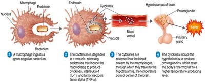
Fever
Systemic response to infection, mediated by cytokines (e.g., IL-1, TNF-α) that reset the hypothalamic thermostat. Benefits include enhanced immune function and inhibition of pathogen growth.
The Complement System
A cascade of serum proteins that enhances immune responses via cytolysis, opsonization, and inflammation. Activated by classical (antibody-dependent), alternative (spontaneous), or lectin (carbohydrate recognition) pathways.
Antimicrobial Substances
Interferons (IFNs): Antiviral proteins produced by infected cells.
Iron-binding proteins: Sequester iron to limit bacterial growth.
Antimicrobial peptides (AMPs): Disrupt microbial membranes and inhibit growth.
Adaptive Immunity: Specific Defenses of the Host
Overview
Adaptive immunity is specific, slower to develop, and has memory. It involves B cells (humoral immunity) and T cells (cellular immunity).
Antigens, Epitopes, and Haptens
Antigens: Substances that trigger immune responses.
Epitopes: Specific regions recognized by antibodies or T cell receptors.
Haptens: Small molecules that require a carrier to be immunogenic.
Antibody Structure and Classes
IgG: Most abundant, crosses placenta, activates complement.
IgM: First produced, pentameric, agglutination.
IgA: Secretions (mucus, milk), mucosal protection.
IgD: On B cells, function unclear.
IgE: Allergic reactions, defense against parasites.
B Cell Activation and Clonal Selection
B cell binds antigen via BCR.
Processes and presents antigen on MHC II.
T helper cell recognizes antigen, releases cytokines.
B cell proliferates into plasma cells (antibodies) and memory B cells.
T Cell Activation and Functions
CD4+ T cells (Helper): Activate B cells and other immune cells.
CD8+ T cells (Cytotoxic): Destroy infected or abnormal cells.
T regulatory cells: Suppress immune responses, prevent autoimmunity.
Immunological Memory
Primary response is slow and weak; secondary response is faster and stronger due to memory cells. Types of immunity include naturally acquired (infection, maternal antibodies) and artificially acquired (vaccination, antibody injection).
Cytokines
Cytokines are protein messengers that regulate immune responses, inflammation, and hematopoiesis. Major types include interleukins, chemokines, interferons, TNF-α, and hematopoietic cytokines.
