 Back
BackComprehensive Study Notes: Viruses, Infectious Disease, and Immunity
Study Guide - Smart Notes
 Tailored notes based on your materials, expanded with key definitions, examples, and context.
Characteristics and Classification of Viruses
General Properties of Viruses
Viruses are acellular, infectious agents composed of nucleic acid (either DNA or RNA) surrounded by a protein coat called a capsid. Some viruses possess an additional phospholipid envelope derived from the host cell membrane. Viruses lack organelles, cytosol, and a cytoplasmic membrane, and are obligate intracellular parasites, meaning they require a host cell for replication.
Virion: The extracellular state of a virus, consisting of the nucleic acid and capsid (nucleocapsid), and sometimes an envelope.
Capsomeres: Protein subunits that make up the capsid.
Envelope: A lipid membrane acquired from the host cell, containing viral proteins and spikes for host recognition and attachment.
Genome: Can be DNA or RNA, single- or double-stranded, linear or circular, but never both DNA and RNA at the same time.
Retroviruses: RNA viruses (e.g., HIV) that carry reverse transcriptase to synthesize DNA from RNA.
Host Range: Viruses infect all forms of life but are usually host-specific.
Size: Viruses range from 10–400 nm and cannot be seen with a light microscope.
Viral Structure and Classification
Classification: Based on nucleic acid type (DNA, RNA, retrovirus), presence of envelope, shape, and size.
Shapes:
Helical: Rod-shaped, e.g., Ebola virus
Polyhedral: Many-sided, most commonly icosahedral, e.g., Adenovirus
Complex: Complicated structures, e.g., T4 bacteriophage
Spikes: Projections from the envelope, often containing enzymes for attachment (e.g., influenza, measles).
Nonenveloped (naked) viruses: Lack an envelope, more resistant to environmental stress.
Enveloped viruses: Have a lipid membrane, more fragile.
Comparison: Bacteria vs. Viruses
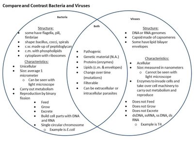
Bacteria are unicellular prokaryotes with metabolic activity, while viruses are acellular and require host cells for replication.
Isolation, Cultivation, and Identification of Viruses
Growing Bacteriophages
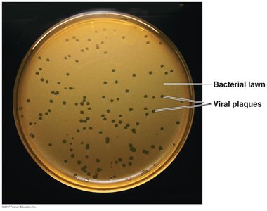
Bacteriophages are grown by infecting bacterial cultures. After infection, the culture is mixed with melted agar and poured into a petri dish. Viral replication leads to the formation of plaques—clear zones where bacteria have been lysed by phages.
Growing Animal Viruses
In living animals: Used to study immune responses.
In embryonated eggs: Viral growth indicated by embryo death or lesions.
In cell cultures: Cells grown in vitro; viral infection leads to cytopathic effects and plaque formation.
Viral Replication
Lytic and Lysogenic Cycles in Bacteriophages
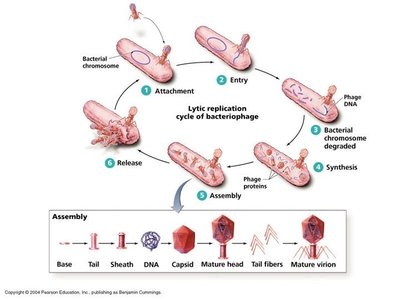
Bacteriophages replicate via two main mechanisms: the lytic cycle (host cell lysis) and the lysogenic cycle (viral DNA integrates into host genome).
Lytic Cycle Steps: Attachment, Entry, Synthesis, Assembly, Release
Lysogenic Cycle Steps: Attachment, Entry, Prophage Integration, Host Cell Replication, Induction, Synthesis, Assembly, Release
Replication of Animal Viruses
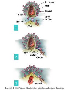
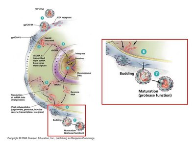
Animal viruses may enter cells by direct penetration, membrane fusion, or phagocytosis. Uncoating removes the capsid, and synthesis depends on the type of nucleic acid. Release occurs by budding (enveloped viruses), exocytosis, or cell lysis.
Latency: Some animal viruses (e.g., herpesviruses, HIV) can remain dormant as proviruses, integrated into host DNA without induction.
Viruses and Cancer
Oncogenic Viruses and Cancer Development
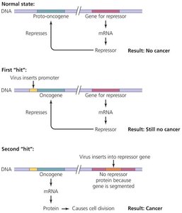
Viruses can contribute to cancer by activating proto-oncogenes or inhibiting tumor suppressor genes. Oncogenic viruses may carry oncogenes, promote host oncogenes, or disrupt tumor repressor genes.
Examples: Hepatitis B virus (liver cancer), Human papillomavirus (cervical cancer)
Viroids and Prions
Viroids
Viroids are small, circular RNA molecules that infect plants, lack a capsid, and do not code for proteins. They cause significant agricultural losses (e.g., potato spindle tuber viroid).
Prions
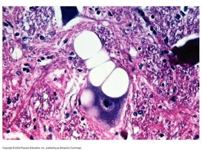
Prions are infectious proteins that lack nucleic acids. They cause disease by inducing misfolding of normal cellular PrP into the pathogenic prion form, leading to neurodegenerative diseases.
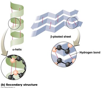
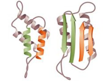
Normal PrP: Alpha-helix structure, functional in brain tissue.
Prion PrP: Beta-pleated sheet structure, forms plaques and vacuoles in neural tissue.
Diseases: Bovine spongiform encephalopathy (BSE), Scrapie, Variant Creutzfeldt-Jakob disease, Kuru, Chronic Wasting Disease.
Transmission: Ingestion, transplantation, or contact with infected tissue.
Prevention: Avoid contaminated meat; prions are resistant to standard sterilization.
Infection, Infectious Disease, and Epidemiology
Symbiosis and Normal Flora
Symbiosis describes the relationship between microorganisms and their hosts, including mutualism, commensalism, and parasitism. Normal microbiota (flora) inhabit various body sites and can inhibit pathogens through competition and production of toxins.
Pathogenicity and Virulence Factors
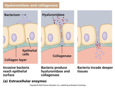
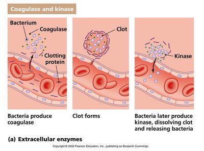
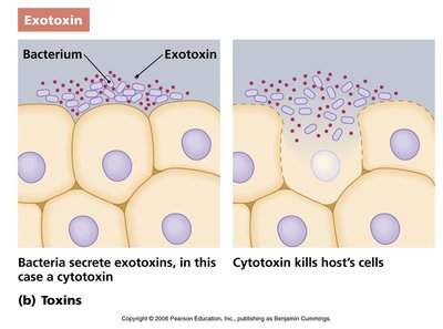
Pathogenicity is the ability to cause disease; virulence is the degree of pathogenicity. Virulence factors include extracellular enzymes, toxins, and antiphagocytic factors.
Extracellular Enzymes: Hyaluronidase, collagenase, coagulase, kinase
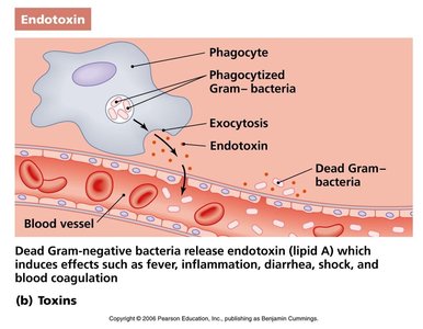
Toxins: Exotoxins (cytotoxins, neurotoxins, enterotoxins) and endotoxins (Lipid A of Gram-negative bacteria)
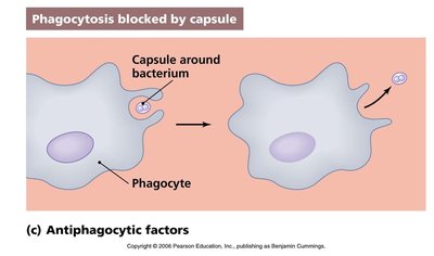
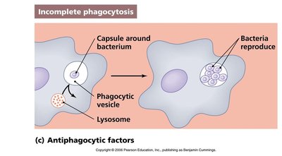
Antiphagocytic Factors: Capsules and chemicals that inhibit phagocytosis
Stages of Infectious Disease
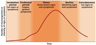
The progression of infectious disease includes incubation, prodromal, illness, decline, and convalescence stages.
Epidemiology and Disease Transmission
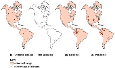
Epidemiology studies the occurrence and spread of disease. Transmission can be direct, indirect, droplet, airborne, waterborne, foodborne, or vector-borne. Disease occurrence is classified as endemic, sporadic, epidemic, or pandemic.
Nosocomial (Healthcare-Associated) Infections
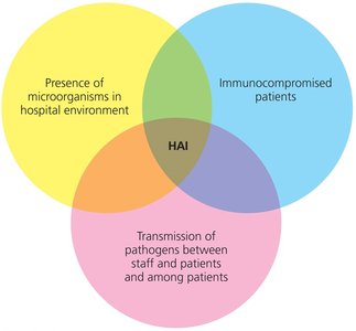
Nosocomial infections are acquired in healthcare settings and are influenced by the presence of microorganisms, immunocompromised patients, and transmission between individuals.
Innate (Nonspecific) Immunity
Lines of Defense
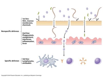
First Line: Physical and chemical barriers (skin, mucous membranes, normal microbiota)
Second Line: Internal defenses (phagocytes, inflammation, fever, antimicrobial substances)
Third Line: Specific (adaptive) immunity (lymphocytes, antibodies)
Phagocytosis
Phagocytes (neutrophils, macrophages, dendritic cells) ingest and destroy pathogens through chemotaxis, adherence (enhanced by opsonins), ingestion, digestion, and elimination.
Fever and Antimicrobial Substances
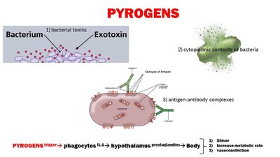
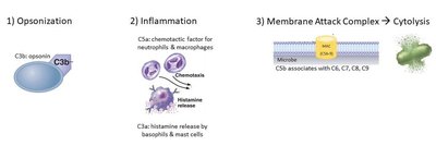
Fever is induced by pyrogens and enhances immune responses. The complement system, interferons, and transferrins are key antimicrobial substances.
Adaptive (Specific) Immunity
Humoral and Cell-Mediated Immunity
Adaptive immunity involves the production of antibodies (humoral) and activation of T cells (cell-mediated) to target specific pathogens. It features immunological memory for faster, stronger responses upon re-exposure.
Lymphatic System and Antigen Presentation
The lymphatic system screens for antigens and facilitates immune responses. Antigen-presenting cells (APCs) process and present antigens via MHC molecules to T cells.
B Cells and Antibodies
B cells produce antibodies (immunoglobulins) specific to antigens. Antibodies neutralize pathogens, activate complement, and enhance phagocytosis.
T Cells and Cytokines
T cells recognize antigens presented by MHC molecules. Cytotoxic T cells kill infected or abnormal cells, while helper T cells coordinate immune responses. Cytokines are signaling molecules that regulate immunity.
Immunological Memory and Acquired Immunity
Memory B and T cells enable rapid secondary responses. Immunity can be naturally or artificially acquired, and active or passive.
Summary Table: Key Differences Between Bacteria and Viruses
Feature | Bacteria | Viruses |
|---|---|---|
Cellular Structure | Prokaryotic cell | Acellular |
Genome | DNA (circular) | DNA or RNA (never both) |
Reproduction | Binary fission | Host-dependent replication |
Metabolism | Present | Absent |
Size | ~1 μm | 10–400 nm |
Pathogenicity | Some pathogenic | Some pathogenic |
