 Back
Control of Microbial Growth and Immune Defense Mechanisms
Study Guide - Smart Notes
 Tailored notes based on your materials, expanded with key definitions, examples, and context.
Controlling Microbial Growth in the Environment
Definitions and Contrasts
Sterilization: The complete destruction or removal of all forms of microbial life, including endospores. Used for surgical instruments, culture media, and pharmaceuticals.
Disinfection: The elimination of most pathogenic microorganisms (except bacterial endospores) on inanimate objects. Used for surfaces and equipment.
Antisepsis: The application of chemical agents to living tissue to inhibit or destroy microorganisms. Used for handwashing and wound cleaning.
Sanitization: Reduction of microbial populations to safe levels as determined by public health standards. Used in food service and public facilities.
Pasteurization: Use of heat to destroy pathogens and reduce spoilage organisms in food and beverages without affecting quality. Commonly used for milk and juices.
Microbial Resistance
Microbes vary in their resistance to control methods. The order from most resistant to most susceptible is:
Bacterial endospores (most resistant)
Mycobacteria
Cysts of protozoa
Vegetative protozoa
Gram-negative bacteria
Fungi
Nonenveloped viruses
Gram-positive bacteria
Enveloped viruses (most susceptible)
Physical Methods of Microbial Control
Moist Heat: Includes boiling, autoclaving, pasteurization, and ultra-high temperature sterilization. Effective for denaturing proteins and destroying membranes.
Dry Heat: Used for sterilizing materials that can withstand high temperatures, such as glassware and metal instruments. Incineration and hot air ovens are common methods.
Lyophilization: Combines freezing and drying to preserve microorganisms for long-term storage.
Ultrafiltration: Used to sterilize heat-sensitive liquids by passing them through a membrane filter that removes microbes based on pore size.
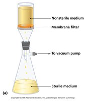
Role of Pore Size in Ultrafiltration: The pore size of the membrane filter determines which microorganisms are removed. Filters with smaller pores can remove bacteria and even some viruses, ensuring sterility of the filtrate.
Radiation:
Non-ionizing (UV light): Causes DNA damage by forming pyrimidine dimers; used for disinfecting air, surfaces, and water.
Ionizing (gamma rays, X-rays, electron beams): Penetrates deeply and causes breaks in DNA; used for sterilizing medical supplies and food.
Biological Monitoring for Sterility
Uses ampules containing Bacillus stearothermophilus spores and a pH indicator.
After autoclaving, ampules are incubated. A color change indicates spore survival and sterilization failure.
If the ampule remains purple, sterilization was successful; yellow indicates failure and the need to redo the load.
Biosafety Levels (BSL)
BSL-1: Basic precautions; non-pathogenic microbes (e.g., Bacillus subtilis).
BSL-2: Moderate risk; pathogens like Staphylococcus aureus.
BSL-3: High risk; airborne pathogens (e.g., Mycobacterium tuberculosis).
BSL-4: Extreme risk; dangerous/exotic agents (e.g., Ebola virus).
Chemical Methods of Microbial Control
Alcohols: Denature proteins and disrupt membranes; used for skin antisepsis and surface disinfection.
Halogens: (Iodine, chlorine, bromine) Oxidize cell components; chlorine is used in water treatment, iodine for skin antisepsis.
Phenolics: Disrupt cell membranes and denature proteins; used in disinfectants.
Aldehydes: (e.g., glutaraldehyde) Cross-link proteins and nucleic acids; used for sterilizing medical equipment.
Gaseous Agents: (e.g., ethylene oxide) Alkylate proteins and DNA; used for heat-sensitive materials.
Oxidizing Agents: (e.g., hydrogen peroxide) Produce free radicals that damage cellular components.
Controlling Microbial Growth in the Body: Antimicrobial Drugs
Modes of Action of Antimicrobial Agents
Inhibition of Cell Wall Synthesis: Prevents formation of peptidoglycan; examples include penicillins and vancomycin.
Inhibition of Protein Synthesis: Targets bacterial ribosomes; examples include tetracycline and aminoglycosides.
Inhibition of Nucleic Acid Synthesis: Inhibits DNA or RNA synthesis; examples include rifampin (RNA) and fluoroquinolones (DNA gyrase).
Disruption of Cell Membrane Function: Damages membrane integrity; examples include polymyxins.
Inhibition of Metabolic Pathways: Antimetabolites block essential enzymes; sulfonamides inhibit folic acid synthesis by competing with PABA.
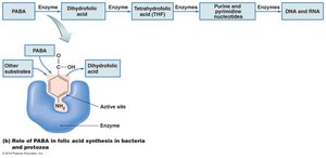
Antimicrobial Susceptibility Testing
Disk Diffusion Method (Kirby Bauer): Measures zones of inhibition around antibiotic disks on agar plates.
Dilution Method (MIC): Determines the minimum inhibitory concentration of an antibiotic that prevents visible growth.
E-Test: Combines aspects of Kirby Bauer and MIC; uses a strip with a gradient of antibiotic concentrations to determine MIC directly on an agar plate.
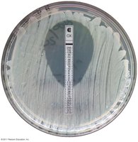
Antimicrobial Resistance
Persister Cells: Dormant variants of regular cells that are tolerant to antibiotics.
Mechanisms of Resistance:
Alteration of cell membrane permeability
Altered drug receptors
Increased drug efflux
Enzymatic inactivation (e.g., beta-lactamase, ESBLs)
Factors Contributing to Resistance: Non-compliance, overuse, and use in agriculture.
Innate and Adaptive Immunity
Innate Immunity (Nonspecific Defense Mechanisms)
First Line of Defense: Skin, mucous membranes, normal microbiota, and secretions (e.g., lysozyme, sebum, antimicrobial peptides).
Second Line of Defense: Phagocytic cells (neutrophils, macrophages), inflammation, fever, and antimicrobial proteins (complement, interferons).
Adaptive Immunity (Specific Defense Mechanisms)
Third Line of Defense: Involves lymphocytes (B cells and T cells) and the production of antibodies.
Antigen Presentation: Antigen-presenting cells (APCs) display antigens to T cells via MHC molecules.
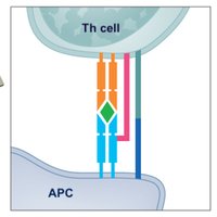
T Cell Activation and Function
Cytotoxic T Cells (CD8+): Eliminate virally infected and tumor cells by recognizing antigens presented with MHC I.
Helper T Cells (CD4+): Regulate immune responses by secreting cytokines and activating B cells and cytotoxic T cells. Th1 cells promote cell-mediated immunity; Th2 cells promote humoral immunity.
B Cell Activation and Antibody Production
T-Dependent Activation: Requires interaction with Th2 cells and antigen presentation via MHC II.
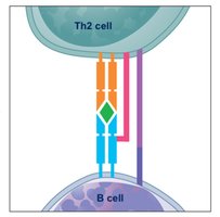
Plasma Cells: Differentiated B cells that secrete antibodies specific to the antigen encountered.
Memory Cells: Long-lived cells that provide rapid response upon re-exposure to the antigen.
Primary and Secondary Immune Responses
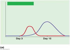
Primary Response: Occurs upon first exposure to an antigen; characterized by a lag period and production of IgM followed by IgG.
Secondary Response: Faster and more robust due to memory cells; predominantly IgG.
Summary Table: Physical and Chemical Methods of Microbial Control
Method | Mechanism | Example/Application |
|---|---|---|
Moist Heat | Denatures proteins, destroys membranes | Autoclaving, boiling |
Dry Heat | Oxidizes cell components | Incineration, hot air oven |
Filtration | Physically removes microbes | Heat-sensitive liquids |
Radiation | Damages DNA | UV for surfaces, gamma for food |
Alcohols | Denature proteins, disrupt membranes | Hand sanitizers |
Halogens | Oxidize cell components | Chlorine in water |
Phenolics | Disrupt membranes, denature proteins | Surface disinfectants |
Aldehydes | Cross-link proteins/DNA | Medical equipment |
Gaseous Agents | Alkylate proteins/DNA | Disposable medical supplies |
Oxidizing Agents | Produce free radicals | Wound cleaning |
Additional info: These notes integrate textbook context and guided reading outlines to provide a comprehensive review for exam preparation in microbiology, focusing on microbial control and immune defense mechanisms.
