 Back
BackCultivation, Isolation, and Identification of Bacteria: Media and Biochemical Tests
Study Guide - Smart Notes
 Tailored notes based on your materials, expanded with key definitions, examples, and context.
Cultivation and Isolation of Bacteria
Fermentation and Oxidation: Key Definitions
Understanding bacterial metabolism is essential for their identification and study. Two fundamental processes are fermentation and oxidation:
Fermentation: An anaerobic metabolic process where bacteria break down sugars (e.g., lactose) to generate energy, producing acidic or gaseous byproducts. This is often detected in the lab by color changes on differential media.
Oxidation: The loss of electrons from a molecule, making it more positive. In contrast, reduction is the gain of electrons, making a molecule more negative.
Application: Fermentation patterns are crucial for identifying enteric pathogens in clinical microbiology.
Pure Culture and Isolation Techniques
To study bacteria accurately, it is necessary to obtain them in pure culture—a state where only one species is present, free from contaminants. This allows for the study of their biochemical, antigenic, and pathogenic properties.
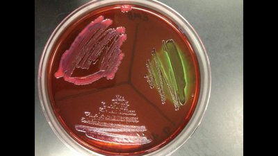
Plating on Solid Media: Clinical samples are streaked onto solid media (e.g., MacConkey, nutrient, or blood agar) to obtain isolated colonies.
Selective Growth Conditions: Use of selective media or anaerobic environments to favor the growth of specific bacteria.
Historical Context: Early methods involved serial dilutions and inoculation into blood. Koch's development of solid media revolutionized isolation, allowing for the separation and identification of individual colonies.
Dilution Series and Quantitative Techniques
A dilution series is used to reduce bacterial concentration stepwise, enabling accurate colony counts and quantification of bacteria in a sample.
Each dilution step reduces the concentration by a factor of 10 (e.g., 1/10, 1/100, 1/1000, etc.).
After plating and incubation, colonies are counted, and the original concentration is calculated using the dilution factor.
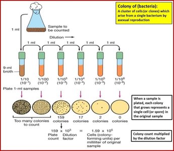
Counting Bacteria: Spread Plate and Pour Plate Methods
These are quantitative techniques for determining the number of viable bacteria in a sample:
Spread Plate: A measured volume of diluted sample is spread evenly on the surface of an agar plate using a sterile spreader.
Pour Plate: Diluted sample is mixed with molten agar and poured into a Petri dish, allowing colonies to grow both on the surface and within the agar.
After incubation, colonies are counted. The ideal range for accurate counting is 25–250 colonies per plate.
Formula for CFU/mL:
c: Concentration (CFU/mL)
n: Number of colonies
d: Dilution factor
s: Volume plated (in mL)
Example: If 159 colonies are counted from a 1:1000 dilution and 1 mL is plated, then:
CFU/mL
Special Media for Bacterial Identification
Types of Media
Selective Media: Contains chemicals that inhibit unwanted bacteria while allowing the desired organism to grow (e.g., Thayer-Martin medium).
Enriched Media: Contains nutrients that enhance the growth of specific bacteria (e.g., Chocolate Agar, Blood Agar).
Differential Media: Contains compounds that allow differentiation of bacteria based on metabolic activity (e.g., MacConkey Agar).
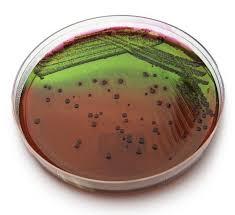
Eosin Methylene Blue (EMB) Agar
EMB agar is both selective and differential, used primarily for Gram-negative enteric bacteria:
Selective: Eosin and methylene blue dyes inhibit Gram-positive bacteria.
Differential: Lactose fermentation produces color changes:
Strong fermenters (e.g., E. coli): Metallic green sheen.
Weak fermenters (e.g., Klebsiella, Enterobacter): Pink to purple colonies.
Non-fermenters (e.g., Salmonella, Shigella): Colorless colonies.
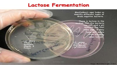
MacConkey Agar
MacConkey agar is selective and differential for Gram-negative enteric bacteria:
Selective: Bile salts and crystal violet inhibit Gram-positive bacteria.
Differential: Lactose fermenters produce pink colonies (due to neutral red pH indicator), while non-fermenters produce colorless or tan colonies.
Examples: E. coli, Klebsiella pneumoniae, and Enterobacter are pink; Salmonella, Shigella, Proteus, and Pseudomonas are colorless.
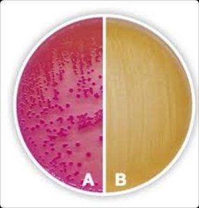
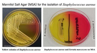
Mannitol Salt Agar (MSA)
MSA is selective and differential, primarily for Staphylococcus species:
Selective: High salt (7.5% NaCl) inhibits most bacteria except halotolerant organisms (mainly Staphylococcus).
Differential: Mannitol fermentation turns the medium yellow (acidic), while non-fermenters keep it red.
Examples: S. aureus (yellow), S. epidermidis (red).
Summary Table: Comparison of Selective and Differential Media
Feature | Mannitol Salt Agar (MSA) | MacConkey Agar (MAC) | Eosin Methylene Blue Agar (EMB) |
|---|---|---|---|
Selectivity | High salt (7.5% NaCl) → only halotolerant organisms (esp. Staphylococcus spp.) grow. | Bile salts & crystal violet → inhibit Gram-positives, allow Gram-negative enterics. | Eosin & methylene blue dyes → inhibit Gram-positives, allow Gram-negative enterics. |
Differentiation | Mannitol + phenol red → mannitol fermenters turn medium yellow, non-fermenters keep it red/pink. | Lactose + neutral red → lactose fermenters form pink/red colonies, non-fermenters remain colorless. | Lactose + dyes → strong lactose fermenters (e.g., E. coli) produce dark purple/black colonies with green metallic sheen; weak fermenters = pink; non-fermenters = colorless. |
Indicator | Phenol red (yellow = acid, red = neutral, pink = alkaline). | Neutral red (red at low pH, colorless at neutral/alkaline). | Eosin & methylene blue (precipitate with acid, giving dark sheen). |
Typical Positive | Staphylococcus aureus (growth + yellow medium). | E. coli, Klebsiella (pink/red colonies). | E. coli (green metallic sheen colonies). |
Typical Negative | Staphylococcus epidermidis (growth but red medium); E. coli inhibited. | Salmonella, Shigella (colorless colonies). | Salmonella, Shigella (colorless colonies). |
Clinical Use | Differentiates pathogenic S. aureus from commensal staphylococci. | Identifies lactose fermenters vs. non-fermenters in enteric infections, UTIs. | Differentiates strong vs weak lactose fermenters in enteric diagnostics, esp. E. coli. |
Biochemical Identification: Fermentation Tests
Lactose, Sucrose, and Glucose Fermentation
Fermentation tests are used to differentiate bacteria based on their ability to ferment specific sugars, producing acid (and sometimes gas):
Lactose Fermentation: Key for distinguishing among Enterobacteriaceae. Positive result = acid/gas production.
Sucrose Fermentation: Useful for differentiating among Enterobacteriaceae.
Glucose Fermentation: All Enterobacteriaceae generally ferment glucose.
Summary Tables: Fermentation Results
Organism | Lactose Fermentation | Sucrose Fermentation | Glucose Fermentation |
|---|---|---|---|
Escherichia | + | + | + |
Klebsiella | + | + | + |
Salmonella | − | + | + |
Shigella | − | − | + |
Proteus | + (some strains) | + | + |
Enterobacter | + | + | + |
Note: '+' indicates fermentation (acid/gas produced); '−' indicates no fermentation.
Colony Morphology on Solid Media
When bacteria are grown on solid media, several characteristics are observed for identification:
Shape: Circular, irregular, radiate, or rhizoid.
Size: Measured in millimeters; useful for identification.
Elevation: The height of the colony above the agar surface.
Margin: Entire, wavy, lobate, filiform.
Surface: Smooth, wavy, rough, granular, papillate, glistening, etc.
Texture: Dry, moist, mucoid, brittle, viscous, butyrous (buttery).
Color: Colorless, pink, black, red, bluish-green, etc.
Summary
This guide covers the essential laboratory techniques for cultivating, isolating, and identifying bacteria, including the use of selective and differential media, quantitative plating methods, and biochemical tests for sugar fermentation. Mastery of these concepts is foundational for microbiology students and critical for clinical diagnostics and research.
