 Back
BackCultivation of Microbes and Control of Microbial Growth
Study Guide - Smart Notes
 Tailored notes based on your materials, expanded with key definitions, examples, and context.
Cultivation of Microbes and Control of Microbial Growth
1. Culturing Microorganisms and Measuring Their Growth
1.1 Culture Media
Culture media are nutrient solutions used to grow microbes in the laboratory. The choice of medium depends on the nutritional requirements of the microorganism and the experimental goals. Media are typically sterilized in an autoclave before use.
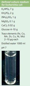
Defined Media: Exact chemical composition is known. Used for precise metabolic studies.
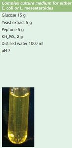
Complex Media: Composed of digests of microbial, animal, or plant products (e.g., yeast and meat extracts). Composition varies slightly from batch to batch.
Enriched Media: Complex media supplemented with highly nutritious substances (e.g., serum, blood) to support the growth of fastidious microorganisms.
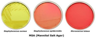
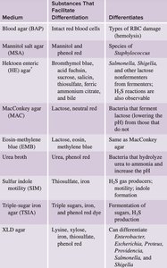
Selective Media: Contains compounds that selectively inhibit the growth of some microbes but not others. Example: Mannitol Salt Agar (MSA) contains high NaCl to inhibit non-halotolerant bacteria.
Differential Media: Contains indicators (dyes or pH indicators) that detect particular metabolic reactions, resulting in a change in colony or media coloration. Example: MSA contains phenol red; fermentation of mannitol changes the color.
1.2 Laboratory Culture
Microbes can be cultured on solid, semi-solid, or liquid media. The physical state of the medium affects the growth pattern and the ability to isolate pure cultures.
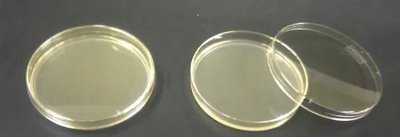
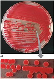
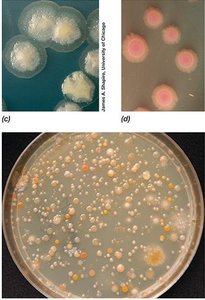
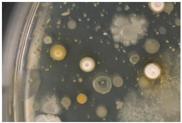
Solid Media: Contains a solidifying agent (e.g., agar). Used in Petri plates and slants. Cells form isolated colonies, each representing a colony-forming unit (CFU).
Semi-solid Media: Contains less agar, used to study bacterial motility.
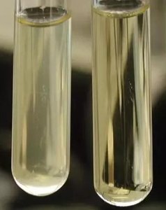
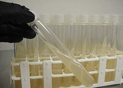
Liquid Media (Broth): No solidifying agent; used in tubes, flasks, or bottles for growing large numbers of cells.
Colony Morphology: Visible characteristics of colonies (size, shape, color, texture, margin, elevation, opacity) are used for identification and to distinguish pure from mixed cultures.
1.3 Quantifying Microbial Growth
Microbial growth can be measured by assessing cell mass or cell number, using direct or indirect methods.
Direct Methods: Wet weight (less accurate), dry weight (more accurate), total nitrogen, quantification of DNA/RNA/proteins/peptidoglycan.
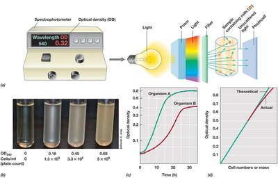
Indirect Methods: Measurement of metabolite consumption/production (e.g., O2, CO2, acid), optical density (OD) via spectrophotometry (turbidity), MacFarland standards.
Standard Curve: OD is proportional to cell number within limits; a standard curve is required for accurate estimation.
1.4 Total Cell Counting
Total cell counting includes both viable (living) and dead cells.
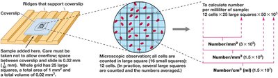
Direct Microscopic Counts: Breed method (staining and counting under a microscope), Petroff–Hausser Counting Chamber (calibrated chamber).
Electron Microscopy: Used for counting viruses.
Coulter Counter: Counts and sizes particles by measuring changes in electrical impedance as particles pass through a small aperture.
1.5 Viable Cell Counting
Viable cell counting estimates the number of living cells capable of forming colonies.
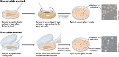
Standard Plate Count: Assumes each bacterium forms a single colony (CFU). Ideal range: 30–300 CFU. Serial dilution may be required.
Spread Plate Method: Bacteria are spread on the surface of agar.
Pour Plate Method: Bacteria are mixed with molten agar and poured into a dish.
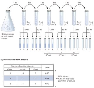
Most Probable Number (MPN): Used for liquid samples; involves dilution series and statistical estimation.
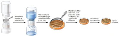
Membrane Filtration: Used for samples with low cell numbers; cells are trapped on a filter and cultured.
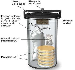
1.6 Culturing Anaerobic Microorganisms
Anaerobic microbes require special conditions to exclude oxygen.
Reducing Media: Chemically remove O2 (e.g., sodium thioglycolate).
Oxygen Indicator: Resazurin indicates the presence of O2.
Anaerobic Jar/Chamber: Used to incubate plates in an oxygen-free environment (e.g., OxyPlate, Oxyrase enzyme).
2. Control of Microbial Growth
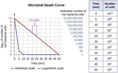
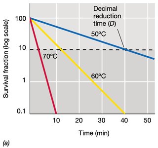
2.1 Microbial Death Curve
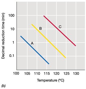
Microbial death occurs at a constant rate, typically following a logarithmic (exponential) decline. The effectiveness of a sterilization or disinfection protocol is often measured by the decimal reduction time (D-value).
Decimal Reduction Time (D-value): Time required to achieve a 1-log (90%) reduction in microbial population at a given condition.
Thermal Death Time: Time to kill all cells at a given temperature.
Thermal Death Point (TDP): Lowest temperature at which all cells in a liquid culture are killed in 10 minutes.
2.2 Physical Agents
Physical methods are widely used to control microbial growth in laboratory and clinical settings.
Temperature (Heat):
Dry Heat: Kills by oxidation (e.g., oven, incineration, flaming).
Moist Heat: Kills by denaturing proteins (e.g., boiling, pasteurization, autoclaving).
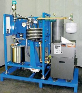
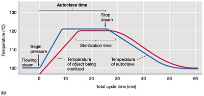
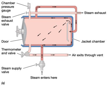
Autoclave: Uses steam under pressure (121°C, 15 PSI, 10–15 min) for sterilization. Steam must contact all surfaces.
Pasteurization: Reduces microbial load but does not sterilize (HTST: 72°C/15 sec; UHT: 138°C/2 sec).
Tyndallization: Repeated heating to kill spores.
Refrigeration and Freezing: Bacteriostatic; slows or stops microbial growth.
Filtration: Used for sterilizing heat-sensitive (thermolabile) solutions. HEPA filters for air; nitrocellulose membrane filters for liquids (pore size 0.2 μm).
Radiation:
UV (270 nm): Non-penetrating, induces DNA mutations (thymine dimers), used for surfaces.
Ionizing Radiation (X-rays, gamma rays): Penetrates tissues, generates reactive oxygen species (ROS), destroys DNA.
D10 Value: Dose required to reduce population by 1 log (90%).

2.3 Chemical Agents
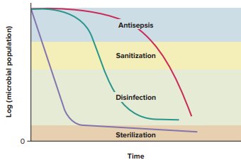
Chemical agents are used to disinfect, sanitize, sterilize, or act as antiseptics. Their effectiveness depends on their chemical structure and mode of action.
Alkylating Agents: Modify DNA, inhibiting replication and protein synthesis.
Surfactants: Disrupt cytoplasmic membranes by dissolving lipids.
Oxidizing Agents: Remove electrons from organic matter, causing cellular damage.
Protein Inactivation: Denaturation, precipitation, or iodination of proteins.
2.4 Effects of Antimicrobial Agents
Antimicrobial agents can be classified by their effect on microbes:
-cidal: Kills microbes (e.g., bactericidal, fungicidal, viricidal). Minimum bactericidal concentration (MBC) is the lowest concentration that kills.
-static: Inhibits growth; removal of agent allows regrowth. Minimum inhibitory concentration (MIC) is the lowest concentration that inhibits growth.
-lytic: Causes cell lysis (e.g., bacteriolytic).
2.5 Effectiveness of Antimicrobial Agents
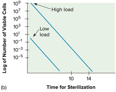
The effectiveness of antimicrobial agents is influenced by several factors:
Population size
Microbial characteristics (e.g., spores, envelopes, Mycobacteria, Gram-positive vs Gram-negative)
Time of exposure
Environmental conditions (pH, temperature, presence of organic matter)
Biofilms
Mode of action and dosage of the agent
Microbial resistance varies, with prions and endospores being most resistant, and enveloped viruses being least resistant.
2.6 Assaying Antimicrobial Activity
Several methods are used to assess the efficacy of antimicrobial agents:
Phenol Coefficient: Historical method comparing effectiveness to phenol (no longer widely used).
Minimum Inhibitory Concentration (MIC): Lowest concentration of an antimicrobial that inhibits visible growth, determined by serial dilution.
Diffusion Method (Kirby-Bauer Test): Discs with antimicrobial agents are placed on inoculated agar; zones of inhibition indicate effectiveness.
Additional info: These notes cover key aspects of microbial cultivation and control, including media types, laboratory techniques, quantification methods, and principles of sterilization and disinfection, as outlined in standard microbiology curricula (Chapters 4 and related sections).
