 Back
BackDiagnostic Clinical Microbiology: Specimen Collection, Processing, and Pathogen Identification
Study Guide - Smart Notes
 Tailored notes based on your materials, expanded with key definitions, examples, and context.
Specimen Collection and Handling
Importance of Proper Specimen Collection
Accurate collection and handling of clinical specimens such as blood, urine, and tissue are foundational to diagnostic microbiology. Proper technique ensures that laboratory results reflect the patient's true condition, minimizing the risk of misdiagnosis and inappropriate treatment.
Accurate Diagnosis: Enables precise identification of infectious agents, guiding effective therapy.
Treatment Monitoring: Allows clinicians to assess patient response to therapy through serial sampling.
Early Detection: Routine screenings can identify infections or risk factors before symptoms develop.
Patient Safety: Prevents errors such as mislabeling or contamination, which can lead to incorrect results and harm.
Tools and Supplies for Specimen Collection
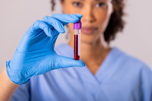
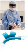
Blood Collection: Utilizes evacuated tubes (e.g., vacutainers), needles, holders, tourniquets, and alcohol wipes for aseptic technique.
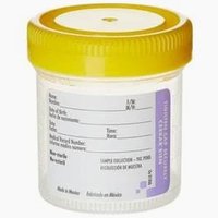
Urine and Other Fluids: Collected in sterile, leak-proof containers with secure lids to prevent contamination and spillage.
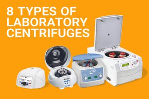
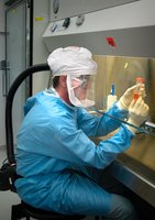
Processing Equipment: Centrifuges are used to separate serum or plasma, while Biosafety Cabinets (BSC) protect personnel during procedures that may generate infectious aerosols.
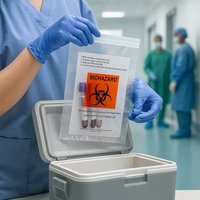
Transport Materials: Include padded carriers, insulated containers with cool packs, and sealed biohazard bags for safe and compliant transport.
Safety Precautions in Clinical Microbiology
Standard Precautions
All human specimens are treated as potentially infectious. Proper use of Personal Protective Equipment (PPE) is mandatory to protect laboratory personnel and prevent cross-contamination.
PPE: Includes gloves, lab coats/gowns, and eye protection.
Aerosol Control: High-risk procedures should be performed in a Biosafety Cabinet to prevent inhalation of infectious particles.
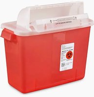
Sharps Safety: Needles must not be bent, broken, or recapped. Immediate disposal in puncture-resistant sharps containers is required.
Spill and Leak Prevention: Secure lids, double-bagging, and decontamination of surfaces with EPA-registered disinfectants are essential.
PPE Maintenance: Gloves must be changed between patients or if damaged. Proper donning and doffing sequences are critical to avoid self-contamination.


Pathogen Identification by Culture and Phenotype
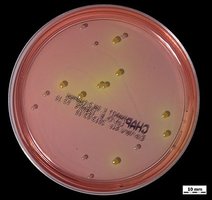
Selective and Differential Media
Diagnostic laboratories use specialized media to isolate and identify pathogens from complex clinical samples. Selective media suppress unwanted microbes, while differential media distinguish organisms based on metabolic traits.
Selective Media: Contain inhibitors (e.g., antibiotics, dyes, high salt) that allow only target organisms to grow. Example: Bile salts or crystal violet inhibit Gram-positive bacteria, aiding in the isolation of Gram-negative pathogens.
Differential Media: Contain indicators that reveal metabolic differences, such as color changes due to acid production. Example: Blood agar differentiates bacteria by hemolysis patterns (beta, alpha, gamma).

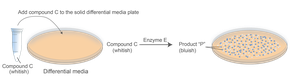
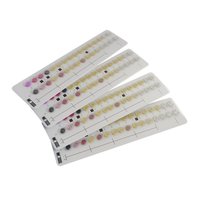
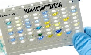
Biochemical Analysis
Biochemical tests identify bacteria based on their enzymatic activities and metabolic pathways. Each species has a unique biochemical fingerprint.
Enzyme Presence: Tests for enzymes like catalase or coagulase.
Substrate Utilization: Determines if bacteria can metabolize specific nutrients (e.g., citrate).
Metabolic By-products: Detects acids or gases produced during metabolism, often indicated by color changes.
Microscopic Analysis in Clinical Microbiology
Core Microscopic Techniques
Microscopy provides rapid, preliminary information about pathogens in clinical samples. Different techniques are chosen based on the organism and required resolution.
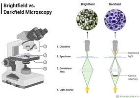
Bright-field Microscopy: Standard method for viewing stained specimens at high magnification (typically 1000x with oil immersion).
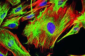
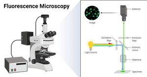
Fluorescence Microscopy: Uses fluorochrome dyes and UV light to detect pathogens at low concentrations.
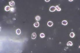
Dark-field Microscopy: Highlights transparent organisms against a dark background, useful for spirochetes like Treponema pallidum.
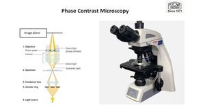
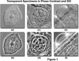
Phase-contrast Microscopy: Enhances contrast in living, unstained cells, ideal for observing motility.
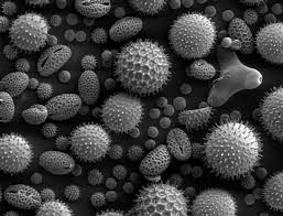
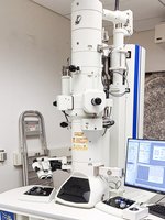
Electron Microscopy (EM): Provides nanometer-scale resolution for visualizing viruses and bacterial ultrastructure.
Clinical Significance of Microscopy
Rapid Results: Provides immediate clues for early treatment decisions.
Quality Control: Detects contaminated or poor-quality samples.
Guided Therapy: Early identification (e.g., Gram-negative diplococci) can direct empiric antibiotic selection.
Molecular Methods for Pathogen Identification
PCR and Nucleic Acid Amplification
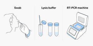
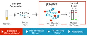
Molecular techniques detect pathogen DNA or RNA, offering speed and sensitivity beyond traditional culture methods.
PCR (Polymerase Chain Reaction): Amplifies specific DNA sequences for detection. Variants include qPCR (quantitative, real-time) and multiplex PCR (multiple targets).
NAAT (Nucleic Acid Amplification Tests): Includes PCR and isothermal methods like LAMP, suitable for point-of-care testing.
Next-Generation Sequencing (NGS): Sequences all genetic material in a sample, useful for identifying unknown or rare pathogens.
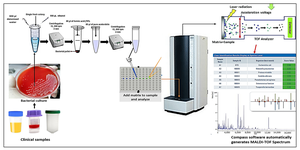
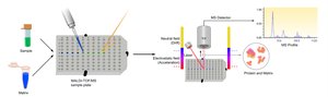
MALDI-TOF MS: Identifies bacteria by their protein mass spectra, providing results in minutes.
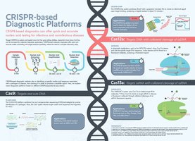
CRISPR-Based Tests: Use CRISPR enzymes to detect specific DNA sequences, producing rapid, visible signals.

Key Advantages of Molecular Methods
Speed: Results in minutes to hours.
Sensitivity: Detects very small amounts of pathogen nucleic acid.
Flexibility: Effective even when pathogens cannot be cultured or are nonviable.
Serological Methods in Diagnostic Microbiology
Principles of Serological Testing
Serological tests detect antibodies produced by the immune system in response to infection, providing evidence of current or past exposure.
IgM: Appears early, indicating recent or current infection.
IgG: Appears later, indicating past infection or immunity.
Both IgM and IgG: Suggest ongoing infection with developing immunity.
Common Serological Tests
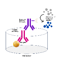
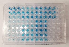
ELISA (Enzyme-Linked Immunosorbent Assay): Detects and quantifies antibodies using a colorimetric change.
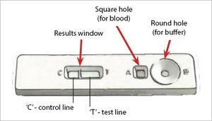
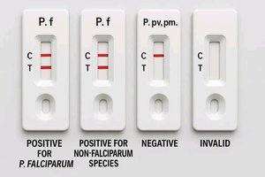
Rapid Diagnostic Tests (RDTs): Provide quick positive/negative results, often in a lateral flow format.
Western Blot: Confirms the presence of specific antibodies with high specificity.
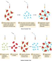
Agglutination Tests: Detect visible clumping when antibodies bind to antigens.
Interpretation of Serological Results
IgM | IgG | Interpretation |
|---|---|---|
+ | − | Recent infection |
+ | + | Recent/ongoing infection |
− | + | Past infection or immunity |
− | − | No exposure or very early infection |
