 Back
Differential Staining Techniques: Acid-Fast and Endospore Stains
Study Guide - Smart Notes
 Tailored notes based on your materials, expanded with key definitions, examples, and context.
Acid-Fast Stain
Principle and Purpose
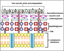
The acid-fast stain is a differential staining technique used to identify bacteria with waxy cell walls containing mycolic acid, which repel aqueous stains. This method is especially important for detecting pathogenic species in the genera Mycobacterium and Nocardia. The stain distinguishes acid-fast organisms from non-acid-fast organisms based on their ability to retain the primary dye after decolorization with acid-alcohol.
Key Point 1: Acid-fast bacteria resist decolorization due to the presence of mycolic acids in their cell walls.
Key Point 2: Non-acid-fast bacteria lose the primary stain and take up the counterstain, appearing a different color.
Example: Mycobacterium tuberculosis is acid-fast, while Staphylococcus epidermidis is non-acid-fast.
Acid-Fast Stain Procedure
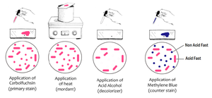
The procedure involves several steps to ensure the primary stain penetrates the waxy cell wall. Heat acts as a mordant to facilitate dye uptake.
Prepare a smear with both acid-fast and non-acid-fast bacteria (e.g., S. epidermidis and M. smegmatis).
Place the slide over steam, cover with paper, and apply carbolfuchsin (primary stain) for 5 minutes, adding stain as needed.
Remove paper, rinse with water.
Decolorize with acid-alcohol until runoff is clear.
Counterstain with methylene blue for 1 minute, rinse with water.
Observe under oil immersion (1000X).
Interpretation of Results
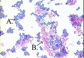
After staining, acid-fast bacteria appear red or pink (retain carbolfuchsin), while non-acid-fast bacteria appear blue (take up methylene blue). Morphology and arrangement can be used to further identify the bacteria.
Key Point: Acid-fast cells = red/pink; Non-acid-fast cells = blue.
Example: In a mixed smear, M. smegmatis (acid-fast rods) will be pink, S. epidermidis (non-acid-fast cocci) will be blue.
Endospore Stain
Principle and Purpose
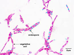
The endospore stain is a differential stain used to visualize bacterial endospores, which are highly resistant, dormant structures formed by certain genera (e.g., Bacillus, Clostridium). Endospores resist most stains, so heat is used to drive the primary stain into the spore.
Key Point 1: Endospores are stained with malachite green and resist decolorization.
Key Point 2: Vegetative cells are decolorized and then counterstained with safranin.
Example: Bacillus subtilis forms endospores that appear green within pink/red vegetative cells.
Endospore Stain Procedure
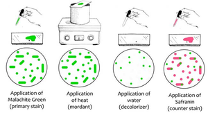
The procedure uses heat to facilitate dye penetration into the endospore, followed by decolorization and counterstaining.
Prepare a smear of the organism.
Place the slide over steam, cover with paper, and apply malachite green (primary stain) for 5 minutes, adding stain as needed.
Remove paper, rinse with water (decolorizer).
Counterstain with safranin for 1 minute, rinse with water.
Observe under oil immersion (1000X).
Interpretation of Results
After staining, endospores appear green, while vegetative cells appear pink/red. This allows for the identification of spore-forming bacteria and assessment of spore location and abundance.
Key Point: Endospores = green; Vegetative cells = pink/red.
Example: Bacillus megaterium endospores can be seen as green ovals within pink rods.
Summary Table: Differential Staining Procedures
Stain | Primary Stain | Mordant | Decolorizer | Counterstain | Result: Target Structure |
|---|---|---|---|---|---|
Acid-Fast | Carbolfuchsin | Heat | Acid-Alcohol | Methylene Blue | Acid-fast cells: pink/red Non-acid-fast: blue |
Endospore | Malachite Green | Heat | Water | Safranin | Endospores: green Vegetative cells: pink/red |
Safety and Disposal
Always wear safety glasses during staining procedures.
Dispose of slides in a sharps container or store in a labeled blue box as directed by your instructor.
Organisms Commonly Used in Lab
Staphylococcus epidermidis (non-acid-fast, non-spore-former)
Mycobacterium smegmatis (acid-fast)
Bacillus subtilis (spore-former; compare 48-hour and 5-day cultures)
Bacillus megaterium (spore-former; compare 48-hour and 5-day cultures)
