 Back
BackDNA and Chromosomes: Structure, Function, and Organization
Study Guide - Smart Notes
 Tailored notes based on your materials, expanded with key definitions, examples, and context.
Chapter 5: DNA and Chromosomes
Part 1: DNA Structure
DNA is the hereditary material in all living organisms, encoding the instructions for life. The discovery of DNA's structure and function was a pivotal moment in biology, leading to our understanding of genetics and molecular biology.
DNA as the Genetic Material
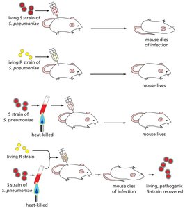
Frederick Griffith's Experiment (1928): Demonstrated the phenomenon of transformation, where heat-killed pathogenic bacteria could transfer virulence to harmless bacteria, suggesting a 'transforming principle.'
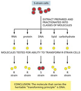
Avery, MacLeod, and McCarty (1944): Identified DNA as the 'transforming principle' responsible for heredity by systematically eliminating other macromolecules.
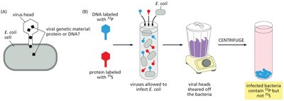
Hershey and Chase (1952): Used bacteriophages labeled with radioactive isotopes to show that DNA, not protein, is the genetic material transmitted to bacteria during infection.
The Structure of DNA
Watson and Crick (1953): Proposed the double helix model of DNA, based on X-ray diffraction data from Rosalind Franklin and Maurice Wilkins.
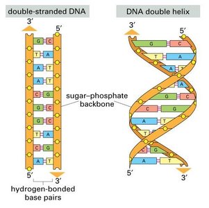
Double Helix: Two antiparallel strands form a right-handed helix with a sugar-phosphate backbone on the outside and nitrogenous bases on the inside.
Base Pairing: Adenine (A) pairs with Thymine (T) via two hydrogen bonds; Guanine (G) pairs with Cytosine (C) via three hydrogen bonds. This complementarity ensures accurate DNA replication.
Helix Properties: Uniform diameter, 10 base pairs per turn, and major/minor grooves for protein interactions.
Nucleotide Structure and DNA Polarity
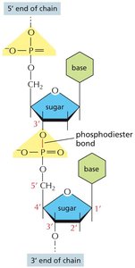
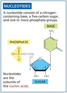
Nucleotides: Each nucleotide consists of a nitrogenous base, a five-carbon sugar (deoxyribose), and one or more phosphate groups.
Phosphodiester Bonds: Link nucleotides together, forming the sugar-phosphate backbone. New nucleotides are always added to the 3' end.
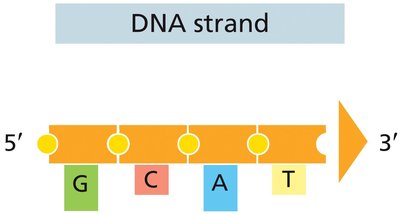
Directionality: DNA strands have polarity, with a free 5' phosphate group at one end and a free 3' hydroxyl group at the other. DNA and RNA are synthesized and read from 5' to 3'.
The Central Dogma of Molecular Biology
The central dogma describes the flow of genetic information: DNA is transcribed into RNA, which is then translated into protein. Some genes encode functional RNAs rather than proteins.
Part 2: The Genome and Chromosomes
The genome is the complete set of genetic material in an organism. In eukaryotes, DNA is organized into chromosomes, which are highly compacted structures composed of DNA and proteins.
Genome Organization
Genome: The total DNA content of an organism. Complexity generally correlates with genome size.
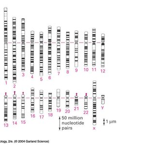
Chromosomes: Long, linear DNA molecules associated with proteins, forming chromatin. Chromosomes are further organized into homologous pairs in diploid organisms.
Chromatin: The complex of DNA and proteins that packages DNA into a compact, organized structure.
Chromosome Anatomy
Centromere: The region where sister chromatids are joined and spindle fibers attach during cell division.
Telomeres: Protective ends of chromosomes that prevent degradation.
Arms: Chromosomes have a short (p) arm and a long (q) arm.
Part 3: DNA Compaction and Organization
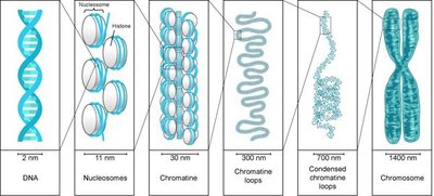
DNA must be compacted to fit within the cell nucleus. This is achieved through multiple levels of organization, primarily involving histone proteins and the formation of nucleosomes.
Levels of DNA Packaging
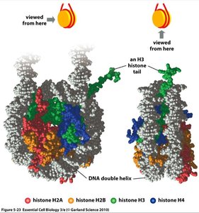
Nucleosome: The basic unit of chromatin, consisting of ~146 base pairs of DNA wrapped around a histone octamer (two each of H2A, H2B, H3, and H4).
30 nm Fiber: Nucleosomes are further packed into a 30 nm fiber, with the help of histone H1.
Higher-Order Structures: The 30 nm fiber forms looped domains, which are further compacted into chromatids during cell division.
Histones and Chromatin Structure
Histones: Small, positively charged proteins rich in lysine and arginine, facilitating interaction with negatively charged DNA.
Histone Modifications: Chemical modifications (e.g., acetylation, methylation) of histone tails can alter chromatin structure and gene expression by changing histone-DNA interactions.
Chromatin Remodeling: Chromatin-remodeling complexes reposition nucleosomes, making DNA accessible for transcription, replication, and repair.
Euchromatin vs. Heterochromatin
Euchromatin: Less condensed, transcriptionally active chromatin.
Heterochromatin: Highly condensed, transcriptionally inactive chromatin. In mammals, one X chromosome in females is inactivated by heterochromatin formation (e.g., calico cats).
DNA Compaction in Eukaryotes vs. Prokaryotes
Eukaryotes: DNA is linear, organized into multiple chromosomes, and packaged with histones into chromatin.
Prokaryotes: DNA is typically circular and not associated with histones, but is compacted by supercoiling and DNA-binding proteins.
Summary Table: Key Features of DNA and Chromosomes
Feature | Description |
|---|---|
Genetic Material | DNA (deoxyribonucleic acid) |
Basic Unit | Nucleotide (base, sugar, phosphate) |
Structure | Double helix, antiparallel strands |
Base Pairing | A-T (2 H-bonds), G-C (3 H-bonds) |
Genome Organization | Chromosomes (linear in eukaryotes, circular in prokaryotes) |
Compaction | Nucleosomes, 30 nm fiber, higher-order structures |
Chromatin Types | Euchromatin (active), Heterochromatin (inactive) |
Additional info: The inactivation of one X chromosome in female mammals (e.g., calico cats) is a classic example of epigenetic regulation via heterochromatin formation. Histone modifications such as acetylation reduce positive charge, loosening DNA-histone interaction and promoting gene expression, while methylation can either activate or repress transcription depending on the context.
