 Back
BackDNA Structure and Replication: Microbiology Study Notes
Study Guide - Smart Notes
 Tailored notes based on your materials, expanded with key definitions, examples, and context.
DNA Structure and Replication
Overview of DNA: Genotype and Phenotype
DNA (deoxyribonucleic acid) is the hereditary material in all living organisms and is responsible for storing genetic information. The genotype refers to the genetic makeup of an organism, while the phenotype is the observable expression of those genes, often as proteins or traits.
Genotype: The sequence of nucleotides in DNA that encodes genetic information.
Phenotype: The physical or biochemical characteristics expressed as a result of gene expression.
Gene Expression: The process by which information from a gene is used to synthesize a functional gene product, typically a protein.
Example: A gene encoding for blue petal color will result in blue petals if expressed.
Structure of DNA
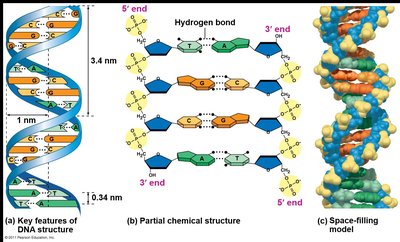
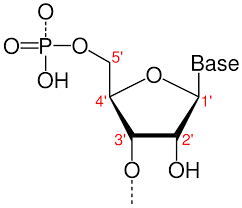
DNA is a double-helical molecule composed of two antiparallel strands. Each strand is a polymer of nucleotides, which consist of a phosphate group, a deoxyribose sugar, and a nitrogenous base.
Nitrogenous Bases: Two types—purines (adenine, guanine) and pyrimidines (cytosine, thymine).
Base Pairing: Adenine (A) pairs with Thymine (T) via two hydrogen bonds; Guanine (G) pairs with Cytosine (C) via three hydrogen bonds.
Backbone: Alternating sugar and phosphate groups connected by phosphodiester bonds.
Antiparallel Strands: One strand runs 5' to 3', the other 3' to 5'.
Ends of DNA: The 5' end has a phosphate group; the 3' end has a hydroxyl group.
Historical Discovery of DNA Structure
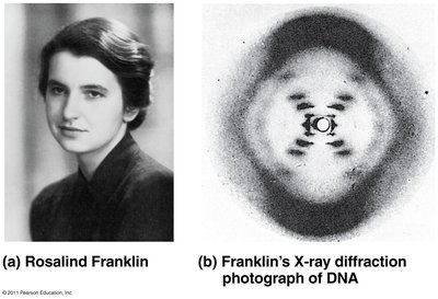
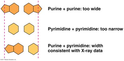
The double helix structure of DNA was elucidated in 1953 by James Watson and Francis Crick, with critical contributions from Rosalind Franklin's X-ray diffraction data.
X-ray Diffraction: Provided evidence for the helical structure of DNA.
Base Pairing: The width of the helix is consistent with purine-pyrimidine pairing.
DNA Supercoiling
Supercoiling refers to the overwinding or underwinding of the DNA double helix. This occurs to compact the DNA so it fits within the cell and to regulate access during replication and transcription.
Function: Facilitates DNA packaging and influences gene expression.
Enzymes: Topoisomerases and gyrases introduce or relieve supercoils.
DNA Replication: General Principles
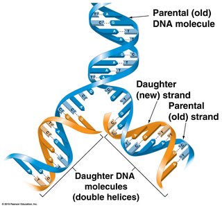
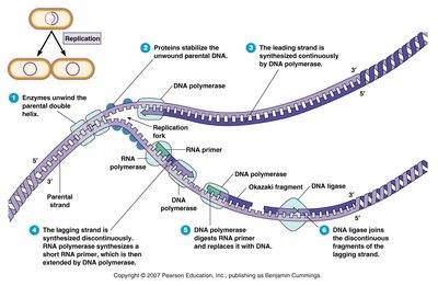
DNA replication is the process by which a cell duplicates its DNA before cell division. It is described as semiconservative because each new DNA molecule consists of one parental and one newly synthesized strand.
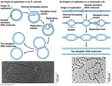
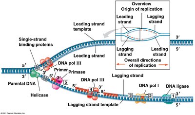
Origin of Replication: Specific sequence where replication begins. Bacteria typically have a single origin; eukaryotes have multiple.
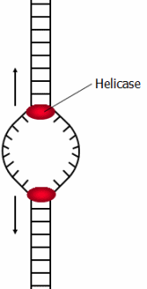
Replication Bubble: The region where the DNA is unwound and replication occurs, containing two replication forks.
Key Enzymes and Proteins in DNA Replication
Several enzymes and proteins coordinate the accurate and efficient replication of DNA:
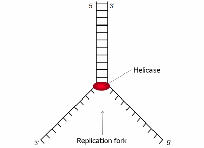
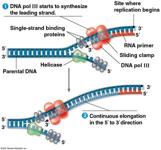
Helicase: Unwinds the parental double helix at the replication fork.
Single-Strand Binding Proteins (SSBs): Stabilize unwound DNA strands and prevent them from re-annealing.
Topoisomerase/Gyrase: Relieves strain ahead of the replication fork caused by unwinding.
Primase: Synthesizes short RNA primers to provide a 3' OH group for DNA polymerase.
DNA Polymerase III: Main enzyme that adds nucleotides to the growing DNA strand in the 5' to 3' direction.
DNA Polymerase I: Removes RNA primers and replaces them with DNA.
DNA Ligase: Joins Okazaki fragments on the lagging strand by forming phosphodiester bonds.
Mechanism of DNA Replication: Leading and Lagging Strands
Because DNA polymerase can only synthesize DNA in the 5' to 3' direction, replication proceeds differently on the two template strands:
Leading Strand: Synthesized continuously toward the replication fork.
Lagging Strand: Synthesized discontinuously away from the replication fork in short segments called Okazaki fragments.
Okazaki Fragments: Short DNA fragments synthesized on the lagging strand, later joined by DNA ligase.
Summary Table: Key Enzymes in DNA Replication
Enzyme/Protein | Function |
|---|---|
Helicase | Unwinds the DNA double helix |
Single-Strand Binding Proteins | Stabilize single-stranded DNA |
Topoisomerase/Gyrase | Relieves supercoiling tension |
Primase | Synthesizes RNA primers |
DNA Polymerase III | Main DNA synthesizing enzyme |
DNA Polymerase I | Removes RNA primers, replaces with DNA |
DNA Ligase | Joins Okazaki fragments |
Importance of Hydrogen Bonds in DNA
Hydrogen bonds between base pairs are weak enough to allow the DNA strands to separate during replication, but strong enough to maintain the double helix structure under normal conditions.
Advantage: Facilitates strand separation during replication and transcription.
Practice Questions and Applications
If a DNA sequence has 5 T and 7 G nucleotides, what will the complementary strand have? Answer: 5 A and 7 C.
Is DNA negatively or positively charged? Answer: DNA is negatively charged due to its phosphate backbone.
Why is DNA replication called semiconservative? Answer: Each new DNA molecule contains one parental and one new strand.
Additional info:
DNA supercoiling is especially important in prokaryotes, which have circular chromosomes, to compact the genome and facilitate replication.
Errors in replication are corrected by proofreading activity of DNA polymerases, ensuring high fidelity of genetic information transfer.
