 Back
BackEnterobacteriaceae and Pseudomonads: Structure, Pathogenesis, and Clinical Relevance
Study Guide - Smart Notes
 Tailored notes based on your materials, expanded with key definitions, examples, and context.
Family Enterobacteriaceae
General Characteristics
The Enterobacteriaceae are a large family of Gram-negative rods, comprising over 50 genera and hundreds of species. Many are enteric (inhabiting the gastrointestinal tract), but most diseases caused by this family occur outside the GI tract. Together with Pseudomonads, they account for about half of all nosocomial (hospital-acquired) infections and are a frequent cause of diarrhea due to their production of enterotoxins.
Facultative anaerobes: Can grow with or without oxygen.
Ferment glucose, oxidase negative, catalase positive.
Divided into coliforms (lactose fermenters) and non-coliforms (non-lactose fermenters).
Some possess peritrichous flagella (flagella distributed over the entire cell).
Identified using enrichment, selective, and differential media.
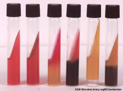
Laboratory Identification
Triple Sugar Iron (TSI) agar is used to differentiate Enterobacteriaceae based on their ability to ferment sugars and produce hydrogen sulfide.
TSI contains glucose (low concentration), lactose, and sucrose (high concentration).
Results are interpreted based on color changes and gas/H2S production.
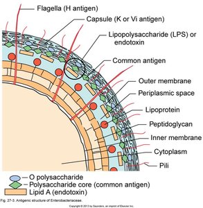
Antigenic Structures
Surface antigens contribute to pathogenicity, trigger immune responses, and are used for serotyping (identification by agglutination):
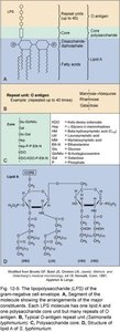
O antigen: Present in all, part of lipopolysaccharide (LPS), acts as an endotoxin (e.g., E. coli O157:H7).
K antigen: Capsule antigen, not present in all species.
H antigen: Flagellar antigen, not present in all species.
Phase variation: Some bacteria can switch expression of K and H antigens on or off.
Virulence Factors
Virulence factors enable Enterobacteriaceae to cause disease:
Endotoxin (Lipid A of LPS): Triggers strong immune responses.
Capsules: Protect against phagocytosis.
Type III secretion systems: Inject bacterial proteins into host cells, manipulating host cell function.
Siderophores: Scavenge iron from the host.
Exotoxins: Various toxins that damage host tissues.
Coliforms and Non-Coliforms
Coliforms
Coliforms are lactose-fermenting Enterobacteriaceae, commonly used as indicators of fecal contamination in water and food. They are generally opportunistic pathogens.
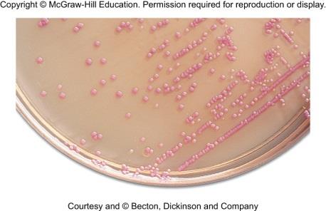
Escherichia coli: Most common aerobic, non-fastidious bacterium in the gut. Pathogenic strains cause diarrhea, UTIs, neonatal meningitis, and more.
Klebsiella pneumoniae: Large capsule, causes pneumonia, meningitis, bacteremia, wound infections, and UTIs. Some strains are carbapenem-resistant (superbugs).
Enterobacter and Citrobacter: Cause UTIs and wound infections.
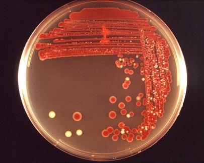
Serratia marcescens: Produces red pigment, causes pneumonia, wound infections, septicemia, and meningitis.
Non-Coliforms
Non-coliforms do not ferment lactose. Many are opportunists, but some are primary pathogens.
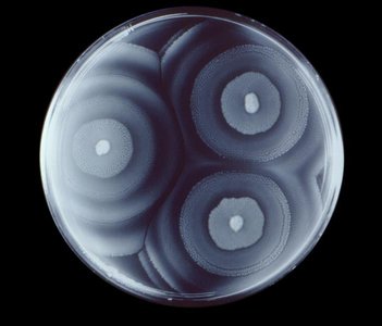
Proteus, Morganella, Providencia: Cause UTIs, wound infections, pneumonia, septicemia, and infant diarrhea. Proteus species swarm on moist agar.
Salmonella and Shigella: Primary pathogens, not part of normal human flora.
Pathogenic Enterobacteriaceae
Escherichia coli Pathotypes
Pathogenic E. coli strains are classified by their virulence factors and clinical syndromes:
ETEC (Enterotoxigenic): Produces heat-labile and heat-stable toxins, causes traveler's diarrhea.
EAEC (Enteroaggregative): Aggregative adherence, stimulates mucus secretion, causes persistent diarrhea.
EIEC (Enteroinvasive): Invades colonic epithelium, causes dysentery-like illness.
EPEC (Enteropathogenic): Destroys microvilli, causes infantile diarrhea.
EHEC (Enterohemorrhagic): Produces Shiga-like toxin, causes hemorrhagic colitis and hemolytic uremic syndrome (e.g., O157:H7).
Clinical relevance: Pathogenic E. coli are a leading cause of infantile diarrhea, traveler's diarrhea, UTIs, and neonatal meningitis.
Other Important Pathogens
Salmonella enterica: Causes typhoid fever and gastroenteritis. Transmission is via contaminated food or water. Chronic carriers may shed bacteria from the gallbladder.
Shigella: Causes bacillary dysentery (shigellosis), characterized by bloody, mucoid diarrhea. Very low infectious dose (ID50 = 50–200 cells).
Yersinia enterocolitica and Y. pseudotuberculosis: Cause enteric infections, can mimic appendicitis.
Yersinia pestis: Agent of plague, transmitted by fleas from wild rodents. Causes bubonic, septicemic, and pneumonic plague.
Clinical Manifestations and Epidemiology
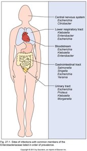
Sites of Infection
Enterobacteriaceae can infect multiple organ systems, including the central nervous system, respiratory tract, bloodstream, gastrointestinal tract, and urinary tract.
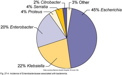
Bacteremia Incidence
The most common Enterobacteriaceae associated with bacteremia are Escherichia, Klebsiella, and Enterobacter.
Genus | Incidence (%) |
|---|---|
Escherichia | 45 |
Klebsiella | 22 |
Enterobacter | 20 |
Proteus | 4 |
Serratia | 4 |
Citrobacter | 2 |
Other | 3 |
Pseudomonads and Related Non-Fermenters
General Characteristics
Pseudomonads are small, monotrichous (single flagellum) Gram-negative rods, primarily found in soil and water. They are important decomposers and bioremediators, but also frequent contaminants in clinical settings. They are notable for their multidrug resistance and opportunistic infections, especially in immunocompromised patients (e.g., cystic fibrosis, burn patients).
Oxidase positive (distinguishes from Enterobacteriaceae).
May produce water-soluble pigments (e.g., pyocyanin).
Common genera: Pseudomonas, Burkholderia, Stenotrophomonas, Acinetobacter, Moraxella.
Pseudomonas aeruginosa
Pseudomonas aeruginosa is a common soil and water organism, found in the intestines of about 10% of people. It is highly resistant to disinfectants, drugs, and drying, and is a frequent contaminant of medical equipment. It is an opportunistic pathogen with a wide range of virulence factors:
Adhesins, flagella, pili, LPS, alginate capsule
Exotoxin A: Blocks protein synthesis in host cells.
Pyocyanin: Greenish-blue pigment, toxic to other microbes and host cells.
Causes endocarditis, meningitis, bronchopneumonia, wound/burn infections.
Characteristic grape-like odor and blue pus in infections.
Other Non-Fermenters
Burkholderia: Respiratory and wound infections; susceptible to trimethoprim/sulfamethoxazole.
Stenotrophomonas: Bacteremia and pneumonia, especially in patients on long-term antibiotics.
Acinetobacter: Survives on dry skin, causes UTIs, respiratory, and wound infections; highly antibiotic-resistant.
Moraxella: Causes otitis and respiratory infections; produces beta-lactamase.
Prevention and Control
Transmission and Prevention
Enterobacteriaceae and Pseudomonads are transmitted via the "5 Fs" (feces, fingers, flies, food, fomites) and water. Prevention includes sanitation, proper food storage, and adequate cooking. When traveling, the "5 Bs" (bread, bananas, beer, bottled beverages, boiled water) are generally safe. The CDC recommends: "Boil it, cook it, peel it, or forget it."
Summary Table: Key Features of Major Enterobacteriaceae
Genus | Lactose Fermentation | Diseases | Key Features |
|---|---|---|---|
Escherichia | Yes | Diarrhea, UTI, meningitis | Many pathotypes, O157:H7, coliform |
Klebsiella | Yes | Pneumonia, UTI, wound | Large capsule, nosocomial |
Enterobacter | Yes | UTI, wound | Opportunist, coliform |
Serratia | Yes | Pneumonia, wound | Red pigment |
Proteus | No | UTI, wound | Swarming motility |
Salmonella | No | Typhoid, gastroenteritis | Primary pathogen |
Shigella | No | Dysentery | Low infectious dose |
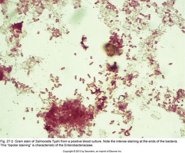
Yersinia | No | Plague, enterocolitis | Bipolar staining, zoonotic |
