 Back
Eukaryotic Cell Structure and Function: Comparison with Prokaryotes
Study Guide - Smart Notes
 Tailored notes based on your materials, expanded with key definitions, examples, and context.
Unique Characteristics of Eukaryotic Cells
Overview of Eukaryotic Cell Structure
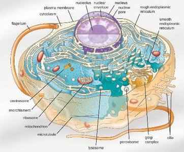
Eukaryotic cells are defined by the presence of a true nucleus and membrane-bound organelles, distinguishing them from prokaryotic cells. Understanding the structure and function of these components is essential for grasping how eukaryotic cells operate as complex living systems.
Nucleus: Contains the cell's genetic material and controls cellular activities.
Membrane-bound organelles: Specialized compartments for distinct cellular processes (e.g., mitochondria, endoplasmic reticulum, Golgi apparatus).
Complex cytoskeleton: Provides structural support and facilitates intracellular transport.
Comparison of Cell Types: Prokaryotes vs. Eukaryotes
Major Differences in Structure and Organization
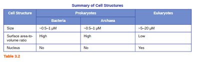
Prokaryotic and eukaryotic cells differ in size, complexity, and internal organization. These differences are fundamental to their biology and evolutionary history.
Prokaryotes: Include Bacteria and Archaea; lack a nucleus and most organelles.
Eukaryotes: Include protists, fungi, plants, and animals; possess a nucleus and various organelles.
Cell Structure | Bacteria | Archaea | Eukaryotes |
|---|---|---|---|
Size | ~0.5–1 μm | ~0.5–1 μm | ~5–20 μm |
Surface area-to-volume ratio | High | High | Low |
Nucleus | No | No | Yes |
Relative Sizes of Biological Structures
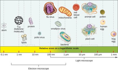
Cells and their components vary greatly in size, which influences their function and the methods used to study them.
Viruses: 20–300 nm
Bacteria: ~1 μm
Eukaryotic cells: 10–100 μm
Eukaryotic Cell Morphologies
Diversity of Eukaryotic Cell Shapes
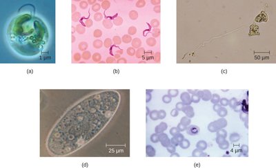
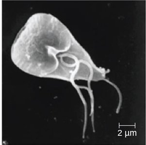
Eukaryotic cells exhibit a wide range of morphologies, reflecting their diverse functions and evolutionary adaptations. Examples include amoeboid, ciliated, and flagellated forms.
External Structures: Flagella and Cilia
Structure and Function
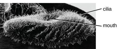
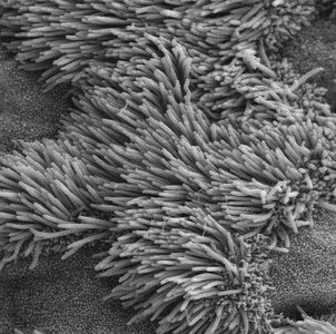
Flagella and cilia are external appendages used for locomotion and movement of substances across the cell surface. They are composed of microtubules arranged in a characteristic "9+2" pattern in eukaryotes.
Flagella: Long, few in number, used for cell movement (e.g., sperm cells).
Cilia: Short, numerous, used for movement or to move substances along the cell surface (e.g., respiratory tract cells).
Composition: Both are made of microtubules (tubulin) and are covered by the plasma membrane.
Surface Structures: Glycocalyx and Cell Wall
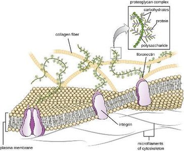
Glycocalyx (Extracellular Matrix)
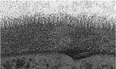
The glycocalyx is a carbohydrate-rich layer outside the plasma membrane, providing protection, cell recognition, and adhesion.
Functions: Protection, cell-cell communication, and anchoring cells within tissues.
Composition: Glycoproteins and glycolipids.
Cell Wall Differences
Most prokaryotes have a cell wall containing peptidoglycan, while eukaryotic cell walls (if present) are chemically distinct and lack peptidoglycan.
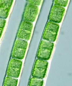
Algae: Cell wall contains cellulose.
Fungi: Cell wall contains chitin.
Animals and protozoa: Lack a cell wall.
Plasma Membrane and Membrane Transport
Structure and Composition
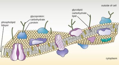
The eukaryotic plasma membrane is a selectively permeable barrier composed of a phospholipid bilayer with embedded proteins, sterols (e.g., cholesterol), and carbohydrates.
Phospholipids: Form the basic structure (bilayer).
Sterols: Provide membrane stability (e.g., cholesterol in animal cells).
Proteins: Serve as receptors, channels, and enzymes.
Membrane Transport Mechanisms
Eukaryotic cells use several mechanisms to transport substances across the plasma membrane:
Simple diffusion: Movement of small, nonpolar molecules down their concentration gradient.
Facilitated diffusion: Movement of molecules via membrane proteins.
Active transport: Movement against the concentration gradient using energy (ATP).
Endocytosis: Uptake of substances by engulfing them in vesicles (includes phagocytosis, pinocytosis, and receptor-mediated endocytosis).
Exocytosis: Release of substances from the cell via vesicle fusion with the plasma membrane.
Cytoplasm and Cytoskeleton
Components and Functions
The cytoplasm consists of cytosol (fluid portion), organelles, and the cytoskeleton. The cytoskeleton provides structural support, maintains cell shape, and facilitates movement of materials within the cell.
Microfilaments: Composed of actin; involved in cell movement and shape changes.
Intermediate filaments: Provide mechanical support; composed of various proteins (e.g., keratin).
Microtubules: Composed of tubulin; involved in cell division, intracellular transport, and structure of cilia/flagella.
Centrosomes are microtubule-organizing centers important for cell division in animal cells.
Internal Structures: Ribosomes and Organelles
Ribosomes
Ribosomes are the sites of protein synthesis. Eukaryotic ribosomes are larger (80S) than prokaryotic ribosomes (70S) and can be free in the cytoplasm or bound to the rough endoplasmic reticulum.
80S ribosome: Composed of a 60S large subunit and a 40S small subunit.
Function: Translate mRNA into proteins.
Nucleus
The nucleus is the control center of the cell, housing the genetic material (DNA) and coordinating activities such as growth, metabolism, and reproduction.
Nuclear envelope: Double membrane with nuclear pores for transport.
Nucleoplasm: Semi-fluid matrix inside the nucleus.
Nucleolus: Site of ribosomal RNA synthesis.
Chromatin: Uncondensed DNA; becomes chromosomes during cell division.
Endomembrane System
The endomembrane system includes the nuclear envelope, endoplasmic reticulum (ER), Golgi apparatus, lysosomes, and vesicles. It is responsible for the synthesis, modification, and transport of proteins and lipids.
Rough ER: Studded with ribosomes; site of protein synthesis and modification.
Smooth ER: Lacks ribosomes; involved in lipid synthesis and detoxification.
Golgi apparatus: Modifies, sorts, and packages proteins and lipids for secretion or delivery to other organelles.
Lysosomes: Contain digestive enzymes for breakdown of macromolecules.
Peroxisomes: Contain enzymes for oxidation reactions and breakdown of hydrogen peroxide.
Vesicles: Transport materials within the cell.
Mitochondria and Chloroplasts
Mitochondria are the sites of aerobic respiration and ATP production. Chloroplasts, found in plants and algae, are the sites of photosynthesis.
Mitochondria: Double-membraned; contain their own DNA and ribosomes.
Chloroplasts: Double-membraned; contain thylakoids, grana, and stroma; also have their own DNA and ribosomes.
Summary Table: Key Differences Between Prokaryotic and Eukaryotic Cells
Feature | Prokaryotes | Eukaryotes |
|---|---|---|
Nucleus | Absent | Present |
Organelles | Absent | Present |
Cell Wall | Usually present (peptidoglycan) | Varies; never peptidoglycan |
Ribosomes | 70S | 80S |
Size | 0.5–1 μm | 5–20 μm |
Flagella | Simple structure | Complex (9+2 microtubules) |
Key Terminology
Extracellular matrix
Cilia and flagella
Sterols
Exocytosis
Endocytosis
Phagocytosis
Pinocytosis
Receptor-mediated endocytosis
Cytoskeleton
Microtubules, intermediate filaments, microfilaments
Centrosome, centrioles
Ribosomes (ribosomal subunits)
Nucleus, nucleolus, nuclear pores, chromatin
Endomembrane system
Rough ER, Smooth ER
Golgi apparatus, cisternae, lumen
Lysosome, peroxisome, vesicles
Mitochondria, chloroplast, thylakoid, granum, stroma
