 Back
Eukaryotic Cells and Microorganisms: Structure, Function, and Diversity
Study Guide - Smart Notes
 Tailored notes based on your materials, expanded with key definitions, examples, and context.
Eukaryotic Cells: Structure and Function
Overview of the Eukaryotic Cell
Eukaryotic cells are complex, membrane-bound cells that evolved from primitive single-celled organisms. Over time, these cells formed colonies, specialized, and eventually gave rise to multicellular organisms. Eukaryotic cells are characterized by compartmentalization of functions into organelles.
Key Features: Presence of a nucleus, membrane-bound organelles, and complex internal structures.
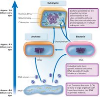
Evolution: Endosymbiosis played a critical role in the development of mitochondria and chloroplasts.
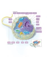
External Structures: Flagella, Cilia, and Glycocalyx
Eukaryotic cells may possess external appendages for movement and interaction with their environment.
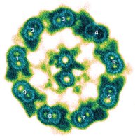
Flagella: Thicker and more complex than bacterial flagella, composed of a 9 + 2 arrangement of microtubules, and covered by the cell membrane.
Cilia: Shorter, more numerous, and structurally similar to flagella; found in protozoa and some animal cells.
Glycocalyx: Outermost layer made of polysaccharides, functioning in protection, adherence, and signal reception.
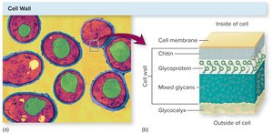
Boundary Structures: Cell Wall and Cell Membrane
The cell wall and membrane provide structural support and regulate interactions with the environment.
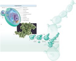
Fungal Cell Walls: Composed of chitin or cellulose (inner layer), glycoproteins, mixed glycans, and an outer glycocalyx. Distinct from bacterial and archaeal cell walls.
Cell Membrane: Phospholipid bilayer with embedded proteins and sterols, providing selective permeability and stability.
Internal Structures and Organelles
The Nucleus
The nucleus is the defining organelle of eukaryotic cells, housing genetic material and coordinating cellular activities.
Nuclear Envelope: Double membrane with pores for molecular exchange.
Nucleolus: Site of ribosomal RNA synthesis and ribosome assembly.
Chromatin: Linear DNA complexed with histone proteins.

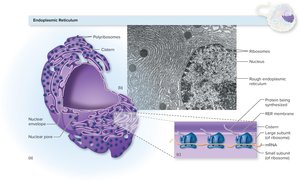
Endoplasmic Reticulum (ER)
The ER is a network of membranes involved in protein and lipid synthesis and transport.
Rough ER (RER): Studded with ribosomes; synthesizes and transports proteins.
Smooth ER (SER): Lacks ribosomes; involved in lipid synthesis and detoxification.
Golgi Apparatus
The Golgi apparatus modifies, sorts, and packages proteins and lipids for secretion or delivery to other organelles.
Structure: Series of flattened sacs (cisternae).
Function: Receives vesicles from the ER, modifies contents, and dispatches them in condensing vesicles.

Vesicular Transport and Lysosomes
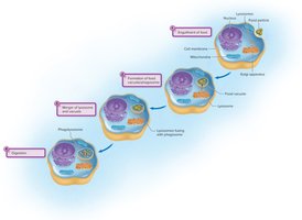
Vesicles mediate transport within the cell, while lysosomes digest macromolecules and cellular debris.
Lysosomes: Contain hydrolytic enzymes for intracellular digestion and defense.
Vacuoles: Store nutrients, waste products, or engulfed particles.

Mitochondria
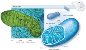
Mitochondria are the energy-generating organelles of eukaryotic cells, responsible for aerobic respiration and ATP production.
Structure: Double membrane with inner folds (cristae) housing enzymes for the electron transport chain.
Unique Features: Contain their own circular DNA and 70S ribosomes, supporting the endosymbiotic theory of origin.
Ribosomes
Ribosomes are the sites of protein synthesis, found free in the cytoplasm, attached to the RER, or within mitochondria and chloroplasts.
Eukaryotic Ribosomes: 80S (60S + 40S subunits).
Bacterial Ribosomes: 70S (found in mitochondria and chloroplasts of eukaryotes).
The Cytoskeleton
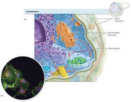
The cytoskeleton provides structural support, facilitates movement, and organizes cellular components.
Actin Filaments: Thin, flexible fibers for cell movement and shape changes.
Intermediate Filaments: Rope-like structures for mechanical strength.
Microtubules: Hollow tubes for organelle movement and cell division.
Endosymbiosis and the Origin of Eukaryotic Organelles
The endosymbiotic theory explains the origin of mitochondria and chloroplasts as descendants of engulfed bacteria, supported by their DNA and ribosome similarities to prokaryotes.
Key Evidence: Circular DNA, independent division, and 70S ribosomes in mitochondria and chloroplasts.
Fungi: Structure, Nutrition, and Reproduction
Fungal Cell Forms and Anatomy
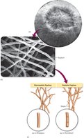
Fungi exist as yeasts (unicellular, round/oval, reproduce by budding) or molds (multicellular, filamentous hyphae). Some are dimorphic, switching forms based on environmental conditions.
Yeasts: Round/oval, reproduce asexually by budding, may form pseudohyphae.
Hyphae: Long, threadlike cells forming the mycelium in molds.
Fungal Nutrition and Ecology
All fungi are heterotrophs, obtaining nutrients from organic substrates. They may be saprobes (decomposers), parasites, or mutualists.
Saprobes: Decompose dead organic matter.
Parasites: Infect living hosts, though most do not require a living host for survival.
Adaptations: Can digest a wide range of substrates and survive in extreme environments.
Fungal Reproduction and Spore Formation
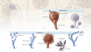
Fungi reproduce by forming spores, either asexually or sexually. Spores are adapted for dispersal and survival in harsh conditions.
Asexual Spores: Sporangiospores (formed in sacs) and conidiospores (free, not enclosed).
Sexual Spores: Result from genetic recombination, increasing diversity.
Protozoa: Structure, Life Cycle, and Pathogenicity
General Characteristics
Protozoa are single-celled eukaryotes with diverse morphologies and life cycles. Most are free-living, but some are important human pathogens.
Cell Structure: Contain all major eukaryotic organelles; cytoplasm divided into ectoplasm and endoplasm.
Locomotion: Move using pseudopods, cilia, or flagella.
Life Cycle: Trophozoite and Cyst Stages
Protozoa often alternate between an active, feeding trophozoite stage and a dormant, resistant cyst stage, aiding survival and transmission.
Trophozoite: Motile, feeding, and reproducing stage.
Cyst: Dormant, resistant to harsh conditions, important for disease transmission.

Major Pathogenic Protozoa
Protozoa are classified by their mode of locomotion and include amoeboid, ciliated, flagellated, and apicomplexan (nonmotile) groups. Each group contains important human pathogens.
Amoeboid: Move by pseudopods (e.g., Entamoeba histolytica).
Ciliated: Move by cilia (e.g., Balantidium coli).
Flagellated: Move by flagella (e.g., Giardia lamblia).
Apicomplexan: Nonmotile, complex life cycles (e.g., Plasmodium species causing malaria).
Helminths: Structure, Life Cycle, and Disease
Major Groups and Morphology
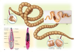
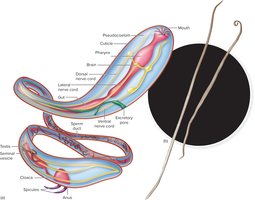
Helminths are multicellular parasitic worms, including flatworms (cestodes and trematodes) and roundworms (nematodes). They have complex organ systems, with the reproductive system being most developed in parasites.
Flatworms: Thin, segmented (tapeworms) or unsegmented (flukes).
Roundworms: Cylindrical, unsegmented, sexes usually separate.
Life Cycle and Transmission
Helminths have complex life cycles involving eggs, larvae, and adults. Transmission often involves intermediate and definitive hosts, with eggs or larvae entering humans via contaminated food, water, or soil.
Egg Laying: High reproductive output ensures survival.
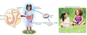
Pinworm Example: Enterobius vermicularis life cycle involves ingestion of eggs, maturation in the intestine, and reinfection via scratching and hand-to-mouth contact.
Summary Table: Key Differences Among Eukaryotic Microorganisms
Group | Cell Type | Motility | Reproduction | Examples |
|---|---|---|---|---|
Fungi | Unicellular (yeasts), multicellular (molds) | Nonmotile | Asexual/sexual spores | Saccharomyces, Aspergillus |
Protozoa | Unicellular | Pseudopods, cilia, flagella | Asexual/sexual | Entamoeba, Giardia |
Helminths | Multicellular | Muscular movement | Sexual (eggs/larvae) | Tapeworms, roundworms |
