 Back
Eukaryotic Cells and Microorganisms: Structure, Function, and Diversity
Study Guide - Smart Notes
 Tailored notes based on your materials, expanded with key definitions, examples, and context.
Chapter 5: Eukaryotic Cells and Microorganisms
The History and Evolution of Eukaryotes
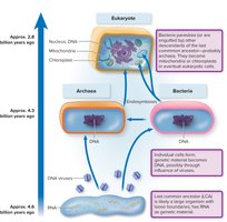
The origin of eukaryotic cells is a fundamental event in the history of life. Eukaryotes are believed to have evolved from a common ancestor that also gave rise to bacteria and archaea. The development of organelles, such as mitochondria and chloroplasts, is explained by the endosymbiotic theory, which proposes that these organelles originated from prokaryotic cells engulfed by ancestral eukaryotes.
Last Common Ancestor (LCA): The precursor cell from which bacteria, archaea, and eukarya diverged.
Endosymbiotic Theory: Suggests mitochondria and chloroplasts were once free-living bacteria that became permanent residents of eukaryotic cells.
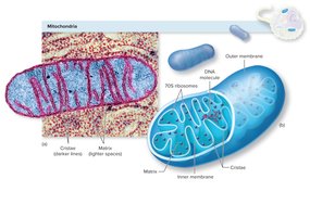
Evidence: Mitochondria and chloroplasts have their own circular DNA, 70S ribosomes, and double membranes.
From Single-Celled to Multicellular Life
Primitive eukaryotes were single-celled organisms. Over time, some cells specialized and formed colonies, eventually leading to multicellular organisms with tissues and organs. This specialization allowed for greater complexity and diversity among eukaryotes.
Structure and Features of Eukaryotic Cells
Generalized Eukaryotic Cell Structure
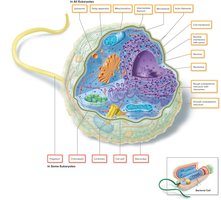
Eukaryotic cells are characterized by the presence of membrane-bound organelles and a true nucleus. All eukaryotic cells share certain structures, while others are found only in specific groups.
Universal Structures: Cytoplasmic membrane, nucleus, mitochondria, endoplasmic reticulum (ER), Golgi apparatus, vacuoles, cytoskeleton, glycocalyx.
Group-Specific Structures: Cell wall, locomotor appendages (flagella, cilia), chloroplasts.
Flagella and Cilia
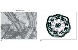
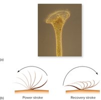
Eukaryotic flagella are structurally more complex and thicker than bacterial flagella. They are covered by the cell membrane and have a characteristic 9+2 arrangement of microtubules, enabling a whipping motion for locomotion. Cilia are similar but shorter and more numerous, functioning in movement and feeding in certain protozoa and animal cells.
Flagella: 9+2 microtubule arrangement, covered by cell membrane, whip-like motion.
Cilia: Short, numerous, oar-like motion, found in protozoa and some animal cells.
The Glycocalyx
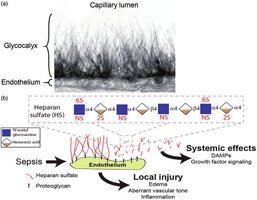
The glycocalyx is the outermost boundary of many eukaryotic cells, composed mainly of polysaccharides. It serves as a protective layer and aids in cell recognition, adhesion, and communication. The glycocalyx can appear as a network of fibers, a slime layer, or a capsule.
Cell Wall
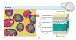
Some eukaryotes, such as fungi and algae, possess a rigid cell wall that provides structural support and shape. Unlike bacterial cell walls, fungal cell walls are composed of chitin or cellulose and various glycoproteins.
Cytoplasmic Membrane
The cytoplasmic membrane is a phospholipid bilayer embedded with proteins and sterols, which stabilize the membrane. It acts as a selectively permeable barrier, regulating the transport of nutrients and waste products.
The Nucleus: The Control Center
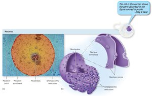
The nucleus is the most prominent organelle, containing the cell's genetic material. It is surrounded by a double-layered nuclear envelope with pores for molecular exchange. The nucleolus within the nucleus is the site of ribosomal RNA synthesis.
Mitosis and Meiosis
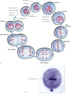
Eukaryotic chromosomes are composed of linear DNA molecules associated with histone proteins. Mitosis is the process of somatic cell division, while meiosis produces sex cells (gametes), ensuring genetic diversity.
Endoplasmic Reticulum (ER)
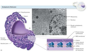
The ER is a network of membranes involved in synthesis and transport. The rough ER (RER) is studded with ribosomes and synthesizes proteins, while the smooth ER (SER) is involved in lipid synthesis and detoxification.
Golgi Apparatus
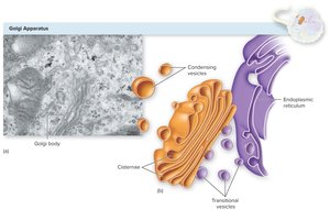
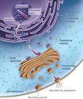
The Golgi apparatus modifies, sorts, and packages proteins and lipids for secretion or delivery to other organelles. It receives transitional vesicles from the ER, processes their contents, and forms condensing vesicles for transport.
Cellular Vesicles: Lysosomes and Vacuoles
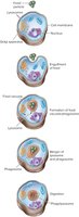
Lysosomes are enzyme-containing vesicles that digest food particles and protect against pathogens. Vacuoles are membrane-bound sacs for storage and transport of substances within the cell.
Mitochondria: Energy Generators
Mitochondria are the sites of aerobic respiration, producing ATP for cellular activities. They have an outer membrane, a highly folded inner membrane (cristae), and a matrix containing DNA and ribosomes. Their bacterial-like features support the endosymbiotic theory.
Ribosomes: Protein Synthesis
Ribosomes are composed of large and small subunits and are responsible for protein synthesis. Eukaryotic ribosomes are 80S (60S + 40S), while mitochondria and chloroplasts contain 70S ribosomes, similar to bacteria.
The Cytoskeleton
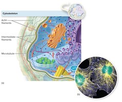
The cytoskeleton provides structural support, facilitates intracellular transport, and enables cell movement. It consists of actin filaments, intermediate filaments, and microtubules.
Fungi: Structure, Nutrition, and Reproduction
Classification and Morphology
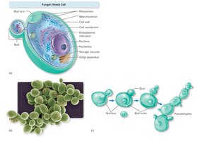
Fungi are divided into macroscopic (mushrooms, puffballs) and microscopic (molds, yeasts) forms. Yeasts are unicellular and reproduce asexually, while molds consist of filamentous hyphae. Some fungi are dimorphic, switching forms based on environmental conditions.
Fungal Nutrition and Organization
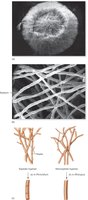
Fungi are heterotrophic, obtaining nutrients from organic matter. Saprobes decompose dead material, while parasites feed on living hosts. Most microscopic fungi form colonies composed of mycelium (a mass of hyphae). Hyphae may be divided by septa, and fungi reproduce via spores.
Functional Types of Hyphae
Hyphae can be vegetative (involved in growth and nutrient absorption) or reproductive (involved in spore production).
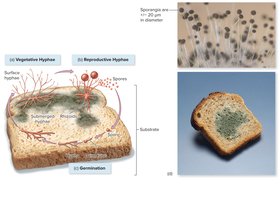
Reproductive Strategies and Spore Formation
Fungi reproduce by outward growth, fragmentation, or spore formation. Asexual spores include sporangiospores (formed in sacs) and conidiospores (free spores). Sexual reproduction introduces genetic variation.
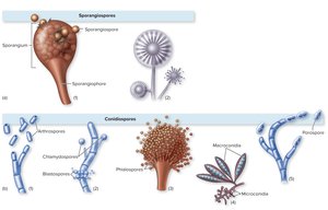
Medical and Beneficial Impacts of Fungi
Pathogenic Fungi: Cause community-acquired, hospital-associated, and opportunistic infections.
Other Impacts: Allergies, toxin-induced neurological conditions, decomposition, symbiosis with plants, and production of antibiotics and food products.
Protists: Algae and Protozoa
General Features
Protists are eukaryotic organisms lacking true tissues. Algae and protozoa are the main subgroups. Protozoa are mostly unicellular, found in diverse habitats, and include both free-living and parasitic species.
Protozoan Structure and Function
Protozoa possess all major eukaryotic organelles except chloroplasts. They have specialized structures for feeding, digestion, and locomotion (pseudopods, flagella, cilia).
Nutrition and Locomotion
Nutrition: Heterotrophic, feeding on organic matter, bacteria, or host fluids/tissues.
Locomotion: Amoeboid motion (pseudopods), flagella, or cilia.
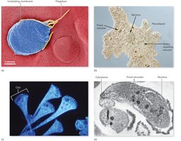
Life Cycle and Reproduction
Protozoa may exist as active trophozoites or dormant cysts. Life cycles can be simple or complex, involving asexual (mitosis, multiple fission) and sexual (conjugation) reproduction. Cyst formation aids survival and disease transmission.
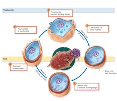
Classification of Protozoa
Flagellates: Move with flagella, form cysts, some are parasitic.
Amoeboids: Move with pseudopods, mostly free-living.
Ciliates: Move with cilia, have complex organelles, mostly harmless.
Sporozoa: Non-motile, complex life cycles, all parasitic.
Important Protozoan Pathogens
Pathogenic protozoa cause diseases such as sleeping sickness (Trypanosoma brucei) and Chagas disease (Trypanosoma cruzi), transmitted by insect vectors.
Helminths: Parasitic Worms
Classification and Morphology
Helminths are multicellular animals, including flatworms (cestodes and trematodes) and roundworms (nematodes). They possess developed organ systems, with the reproductive tract being most prominent. Protective cuticles and specialized mouthparts aid in parasitism.
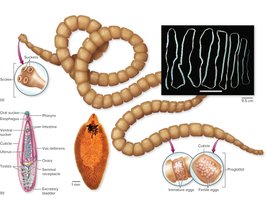
Life Cycles and Transmission
Helminth life cycles include egg, larval, and adult stages. Most derive nutrients and reproduce sexually within the host. Transmission occurs via ingestion of eggs or larvae, or through vector organisms.
Summary Table: Major Elements of Life in Each Organism Type
Feature | Bacteria/Archaea | Eukaryotes | Viruses |
|---|---|---|---|
Cell Type | Prokaryotic | Eukaryotic | Non-cellular |
Organelles | Absent | Present | Absent |
Genetic Material | Circular DNA | Linear DNA in nucleus | DNA or RNA |
Ribosomes | 70S | 80S (70S in organelles) | Absent |
Cell Wall | Peptidoglycan (bacteria) | Chitin/cellulose (fungi/algae) | Absent |
Reproduction | Asexual (binary fission) | Asexual/sexual | Requires host cell |
