 Back
BackEukaryotic Microbes and Invertebrate Infectious Agents: Structure, Function, and Disease
Study Guide - Smart Notes
 Tailored notes based on your materials, expanded with key definitions, examples, and context.
Eukaryotic Microbes
Core Characteristics of Eukaryotic Microbes
Eukaryotic microbes are unicellular or multicellular organisms with complex cellular structures, including a nucleus and membrane-bound organelles. They are distinguished from prokaryotes by their size, complexity, and cellular division mechanisms.
Nucleus: DNA is enclosed within a nuclear membrane.
Organelles: Specialized structures such as mitochondria (energy production) and, in some cases, chloroplasts (photosynthesis).
Larger and More Complex: Eukaryotic cells are typically larger and structurally more intricate than prokaryotic cells.
Cell Division: Eukaryotes divide by mitosis, ensuring accurate chromosome segregation.
Main Groups of Eukaryotic Microbes
Eukaryotic microbes are classified into four primary categories, each with unique biological roles and characteristics.
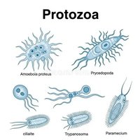
Protozoa: Unicellular, motile organisms often found in aquatic environments. Examples include Amoeba, Paramecium, and Trypanosoma.
Algae: Photosynthetic organisms, ranging from unicellular to multicellular forms, crucial for oxygen production and aquatic food webs.
Microscopic Fungi: Includes yeasts and molds, important for decomposition and biotechnology.
Other Eukaryotic Microbes: Includes slime molds and water molds.


Importance of Eukaryotic Microbes
Eukaryotic microbes play essential roles in ecology, human health, and biotechnology.
Ecological Balance: Algae produce oxygen and form the base of aquatic food chains.
Decomposition: Fungi recycle nutrients by breaking down organic matter.
Human Health: Some cause diseases (e.g., malaria, giardiasis), while others are beneficial (e.g., yeast in bread-making).
Biotechnology: Used in medicine, biofuel production, and food fermentation.

Invertebrate Infectious Agents
Definition and Types
Invertebrate infectious agents are animals without a backbone that either directly cause disease or act as vectors transmitting pathogens.
Direct Parasitic Agents: Helminths (worms), ectoparasites (lice, fleas, mites), and mollusks (snails as hosts for flukes).
Vectors of Infection: Arthropods (mosquitoes, ticks, flies) transmit pathogens; mechanical vectors (houseflies, cockroaches) carry pathogens without internal development.
Pathogens of Invertebrates: Microbes infecting invertebrates, important in biological pest control.

Parasitic Flagellated Protozoa: Genus Giardia
Giardia: Structure and Life Cycle
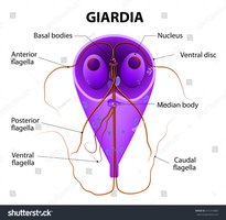
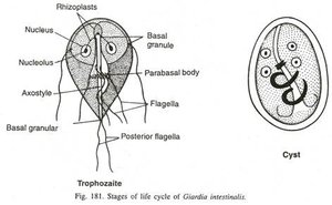
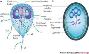
Giardia is a flagellated protozoan that infects the small intestine, causing giardiasis. It alternates between two forms: trophozoite and cyst.
Trophozoite: Pear-shaped, two nuclei, four pairs of flagella, ventral disc for attachment.
Cyst: Oval, thick wall, four nuclei, resistant to environmental stress.
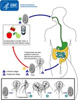
Giardia duodenalis: Infection and Life Cycle
Giardia duodenalis infects humans and animals via ingestion of cysts from contaminated water, food, or surfaces. Excystation releases trophozoites in the small intestine, which multiply and eventually encyst for excretion.
Transmission: Fecal-oral route via cysts.
Excystation: Each cyst releases two trophozoites in the small intestine.
Multiplication: Trophozoites multiply by binary fission.
Encystation: Trophozoites form cysts before excretion.


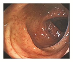
Pathology of Giardiasis
Giardiasis causes gastrointestinal symptoms, including diarrhea, malabsorption, and weight loss. Trophozoites disrupt intestinal epithelial junctions and brush border enzymes, affecting motility and nutrient absorption.
Symptoms: Severe diarrhea, malnutrition, weight loss, epithelial injury.
Mechanism: Adherence via ventral disc, disruption of cell junctions.
Diagnosis of Giardiasis
Diagnosis relies on stool antigen detection assays, nucleic acid amplification tests (NAAT), and microscopy. Multiple stool samples increase sensitivity due to intermittent shedding.
Antigen Detection: Sensitive and specific.
NAAT: Highly sensitive molecular method.
Microscopy: Direct visualization of trophozoites/cysts; sensitivity improved with multiple samples.

Treatment of Giardiasis
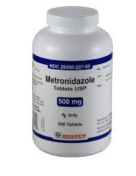
Initial management includes oral rehydration and, if necessary, intravenous fluids. Metronidazole is the first-line drug, with alternatives for special populations.
Metronidazole: First-line therapy; caution in pregnancy.
Other Regimens: Tinidazole, nitazoxanide, mebendazole, albendazole, paromomycin.
Epidemiology of Giardiasis
Giardia is globally distributed and is the most common intestinal parasitic disease in the United States. Risk factors include childcare settings, travel to areas with poor sanitation, recreational water exposure, and contact with infected animals.
Prevalence: Over 1 million cases per year in the U.S.
Risk Factors: Childcare, travel, swimming, animal contact.

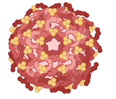
Zika Virus: Structure, Transmission, and Pathogenesis
Structure and Classification
Zika virus is a single-stranded, positive-sense RNA virus in the Flaviviridae family. Other flaviviruses include dengue, yellow fever, and West Nile virus.
Family: Flaviviridae
Genome: Single-stranded, positive-sense RNA
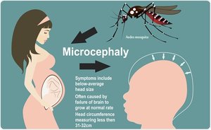
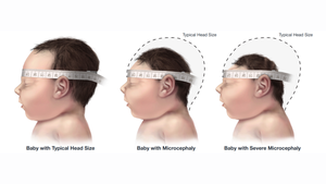
Zika Virus Pathogenesis
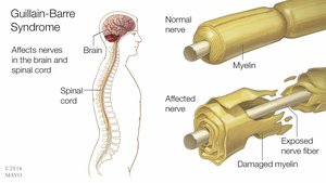
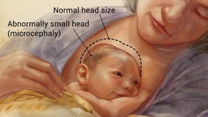
Zika virus primarily affects fetal brain development, causing microcephaly and other neurological defects. In adults, it is associated with Guillain-Barré Syndrome (GBS).
Microcephaly: Destruction of neural progenitor cells leads to reduced brain size and cortical thinning.
GBS: Immune-mediated nerve damage, muscle weakness, and paralysis.
Zika Virus Transmission
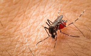
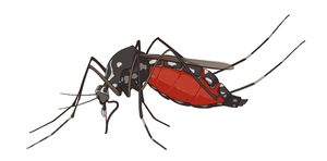
Zika virus is transmitted by Aedes mosquitoes, mother-to-fetus, sexual contact, and rarely by blood transfusion.
Mosquito Bites: Aedes aegypti and Aedes albopictus are primary vectors.
Mother to Fetus: Vertical transmission during pregnancy.
Sexual Contact: Virus persists in semen; transmission possible even if asymptomatic.
Blood Transfusion: Rare, but documented.

Zika Virus Transmission Cycles
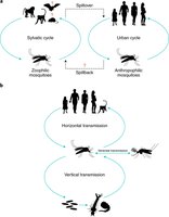
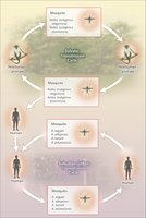
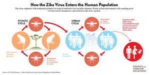
Zika virus transmission occurs in two main cycles: sylvatic (rural/enzootic) and urban. Each cycle involves different hosts and vectors.
Sylvatic Cycle: Forest mosquitoes transmit virus between non-human primates; humans are incidental hosts.
Urban Cycle: Urban mosquitoes transmit virus between humans, especially in densely populated areas.

Zika Virus Pathogenesis and Tissue Tropism
Zika virus exhibits tissue tropism, affecting multiple organs and fluids, including the brain, placenta, eye, testis, uterus, and body fluids.
Brain: Neural progenitor cells, mature neurons, astrocytes.
Placenta: Trophoblasts, endothelial cells.
Eye: Ganglion cells, optic nerve.
Testis: Leydig and Sertoli cells.
Uterus/Vagina: Vaginal epithelial cells.
Body Fluids: Saliva, semen, urine.
Vectors of Zika Virus
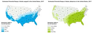
Zika virus is primarily transmitted by Aedes species mosquitoes, with Aedes aegypti as the primary vector and Aedes albopictus as a secondary vector.
Aedes aegypti: Main vector, breeds in domestic water containers.
Aedes albopictus: Secondary vector, also capable of transmission.
Review Questions
Parasitic flagellated protozoa that possess a kinetoplast are primarily classified under which group? Answer: C. Kinetoplastida
Which of the following diseases is caused by a kinetoplastid protozoan transmitted by an insect vector? Answer: C. African sleeping sickness
Which distinguishing feature is characteristic of kinetoplastid protozoa but not Giardia or Trichomonas? Answer: C. Presence of a kinetoplast containing mitochondrial DNA
Zika virus belongs to which family? Answer: B. Flaviviridae
The main mosquito vector of Zika virus is: Answer: C. Aedes aegypti
In adults, Zika virus infection has been associated with: Answer: B. Guillain-Barré Syndrome
The primary mode of Zika virus transmission is: Answer: B. Bite of infected Aedes mosquitoes
Which of the following can transmit Zika virus sexually? Answer: C. Even asymptomatic individuals
The sylvatic cycle of Zika virus involves transmission between: Answer: B. Non-human primates and forest mosquitoes
