 Back
BackEukaryotic Microbes and Invertebrate Infectious Agents: Structure, Function, and Disease
Study Guide - Smart Notes
 Tailored notes based on your materials, expanded with key definitions, examples, and context.
Eukaryotic Microbes
Core Characteristics of Eukaryotic Microbes
Eukaryotic microbes are unicellular or simple multicellular organisms with complex cell structures. They are distinguished from prokaryotes by the presence of a nucleus and membrane-bound organelles.
Nucleus: Contains the cell's genetic material enclosed within a nuclear membrane.
Organelles: Specialized structures such as mitochondria (for energy production) and, in some, chloroplasts (for photosynthesis).
Larger and More Complex: Eukaryotic cells are generally larger and structurally more complex than prokaryotic cells.
Cell Division: Eukaryotic microbes divide by mitosis, ensuring accurate chromosome segregation.
Main Groups of Eukaryotic Microbes
Eukaryotic microbes are classified into four primary categories based on their structure and function:
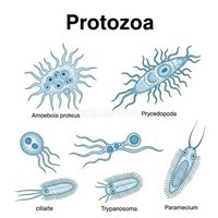
Protozoa: Unicellular, motile organisms, often found in aquatic environments. Examples include Amoeba, Paramecium, and Trypanosoma.
Algae: Photosynthetic organisms, ranging from unicellular to multicellular forms. They are crucial for oxygen production and aquatic food webs.
Fungi: Includes yeasts, molds, and mushrooms. Fungi are decomposers and play a role in nutrient cycling.
Microscopic Helminths: Some multicellular worms are studied in microbiology due to their parasitic nature.


Importance of Eukaryotic Microbes
Ecological Balance: Algae produce a significant portion of Earth's oxygen and are foundational to aquatic food chains.
Decomposition: Fungi decompose organic matter, recycling nutrients in ecosystems.
Human Health: Some eukaryotic microbes are beneficial (e.g., yeast in baking), while others are pathogenic (e.g., Plasmodium causes malaria).
Biotechnology: Used in the production of medicines, biofuels, and fermented foods.
Invertebrate Infectious Agents
Definition and Roles
Invertebrate infectious agents are animals without backbones that can cause or transmit diseases. They may act as direct parasites or as vectors for other pathogens.
1. Direct Parasitic Agents
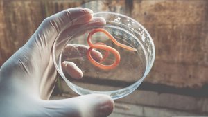
Helminths (Worms): Multicellular parasites such as tapeworms, roundworms, and flukes that infect various tissues.
Ectoparasites: Insects and arachnids like lice, fleas, and mites that live on the host's surface.
Mollusks: Some snails serve as intermediate hosts for parasitic flukes (e.g., schistosomiasis).
2. Vectors of Infection
Arthropod Vectors: Insects and ticks that transmit pathogens, such as mosquitoes (malaria, Zika), ticks (Lyme disease), and flies (sleeping sickness).
Mechanical Vectors: Invertebrates like houseflies and cockroaches that physically transfer pathogens without the pathogen developing inside them.

3. Pathogens of Invertebrates
Invertebrates can also be infected by microbes (viruses, bacteria, fungi), which are important in biological control of pests.
Flagellated Protozoa: Giardia
Genus Giardia
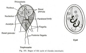
Giardia is a flagellated protozoan parasite that infects the small intestine of vertebrates, causing giardiasis. It alternates between two forms: trophozoite (active, feeding stage) and cyst (infectious, resistant stage).
Life Cycle and Morphology
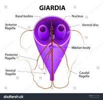
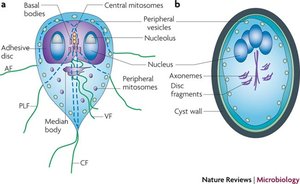
Trophozoite: Pear-shaped, two nuclei, four pairs of flagella, ventral disc for attachment to intestinal mucosa.
Cyst: Oval, thick wall, four nuclei, resistant to environmental stress, formed in the large intestine.
Giardia duodenalis (G. lamblia, G. intestinalis)
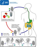
This species infects a wide range of hosts, including humans and animals. Infection occurs via ingestion of cysts in contaminated water, food, or surfaces.
Life Cycle
Cysts are ingested and pass through the digestive tract.
Excystation in the small intestine releases trophozoites.
Trophozoites multiply by binary fission and attach to the intestinal lining.
Encystation occurs as trophozoites move toward the colon; cysts are excreted in feces and remain infectious in the environment.
Pathology
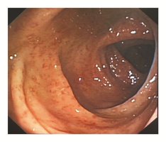
Symptoms: Severe diarrhea, malabsorption, weight loss, and mild intestinal injury.
Mechanism: Trophozoites disrupt epithelial cell junctions and brush border enzymes, altering gastrointestinal motility.
Diagnosis
Stool antigen detection assays and nucleic acid amplification tests (NAAT) are more sensitive than microscopy.
Microscopy sensitivity increases with multiple stool samples.
Microscopy should be performed even if antigen or NAAT tests are used.

Treatment
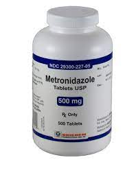
Oral rehydration and IV fluids for dehydration.
Metronidazole is the first-line drug; alternatives include tinidazole, nitazoxanide, mebendazole, albendazole, and paromomycin.
Epidemiology
Giardia is found worldwide and is the most common intestinal parasitic infection in the U.S.
Risk factors: Childcare settings, poor sanitation, recreational water exposure, contact with infected animals.

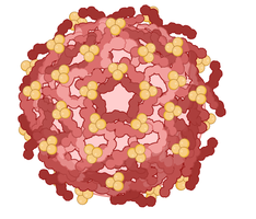
Zika Virus
Classification and Structure
Genus: Orthoflavivirus
Family: Flaviviridae
Genome: Single-stranded, positive-sense RNA
Related Viruses: Dengue, Yellow fever, West Nile virus
Pathogenesis and Clinical Manifestations
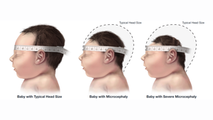
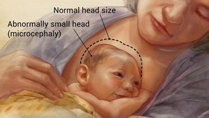
Fetal Brain Development: Zika virus targets neural progenitor cells, causing microcephaly and neurological defects in fetuses.
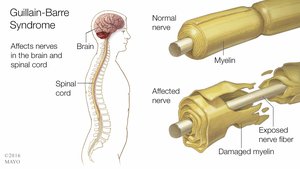
Guillain-Barré Syndrome (GBS): In adults, Zika infection can trigger GBS, leading to muscle weakness and paralysis.
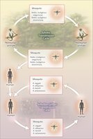
Transmission
Mosquito Bites: Main vectors are Aedes aegypti and Aedes albopictus.
Mother to Fetus: Vertical transmission during pregnancy or birth.
Sexual Contact: Virus can be transmitted sexually, even by asymptomatic individuals.
Blood Transfusion: Rare, but possible.
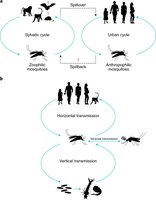
Transmission Cycles
Sylvatic (Rural/Enzootic) Cycle: Involves non-human primates and forest mosquitoes; humans are incidental hosts.
Urban Cycle: Involves human-mosquito-human transmission in densely populated areas.
Vectors
Primary Vector: Aedes aegypti
Secondary Vector: Aedes albopictus

Pathogenesis and Tissue Tropism
Zika virus exhibits tropism for neural, placental, ocular, and reproductive tissues, explaining its diverse clinical manifestations.
Summary Table: Key Features of Giardia and Zika Virus
Feature | Giardia | Zika Virus |
|---|---|---|
Type | Flagellated protozoan | RNA virus (Flaviviridae) |
Transmission | Fecal-oral (cysts in water/food) | Mosquito bite, sexual, vertical, blood transfusion |
Main Disease | Giardiasis (diarrhea, malabsorption) | Zika fever, microcephaly, GBS |
Diagnosis | Stool antigen, NAAT, microscopy | RT-PCR, serology |
Treatment | Metronidazole, rehydration | Supportive (no specific antiviral) |
