 Back
BackEukaryotic Microorganisms: Protozoa, Helminths, and Fungi (chapter 5)
Study Guide - Smart Notes
 Tailored notes based on your materials, expanded with key definitions, examples, and context.
The Eukaryotes of Microbiology
Overview of Eukaryotic Microorganisms
This chapter introduces three major groups of eukaryotic microorganisms: protozoa, helminths, and fungi. These organisms are significant in microbiology due to their roles in human health, disease, and ecological processes. Eukaryotic microorganisms possess complex cellular structures and diverse life cycles, distinguishing them from prokaryotes.
Protozoa: "First Animals"
General Characteristics
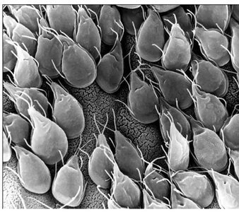
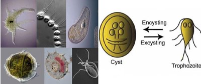
Protozoa are unicellular, nonphotosynthetic, and motile organisms. Many are parasitic, completing their life cycle within a host and potentially causing illness. Protozoa can form cysts through encystment, a survival mechanism during unfavorable conditions. The trophozoite is the feeding and growth stage, while the cyst is a dormant, protective form.
Unicellular and motile
Parasitic species cause human diseases
Encystment: Formation of cysts for survival
Trophozoite: Active, feeding stage
Major Groups of Protozoa
Protozoa are classified into several groups based on their morphology and mode of locomotion:
Excavata: Flagellated protozoa (e.g., Giardia)
Amoebozoa: Ameboid protozoa (e.g., Entamoeba), move via pseudopodia
Chromalveolata: Includes ciliated (Balantidium) and apicomplexan (Plasmodium) protozoa
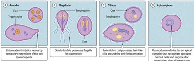
Apicomplexa
Apicomplexan protozoa are obligate parasites, characterized by an apical complex used for host cell attachment and invasion. Their life cycles are complex, often involving multiple hosts and stages. Plasmodium, the causative agent of malaria, is a notable example.
Apical complex: Specialized structure for infection
Sporozoite: Infective stage
Schizogony: Asexual reproduction producing merozoites
Plasmodium: Life cycle involves human and mosquito hosts
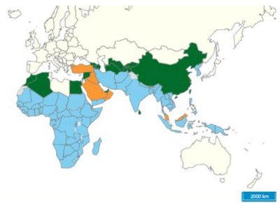
Malaria: Epidemiology and Symptoms
Malaria is a major global health concern, caused by Plasmodium species. Transmission occurs via mosquito bites, blood transfusions, and from mother to fetus. Symptoms include chills, fever, headache, and fatigue, resulting from the rupture of red blood cells by the parasite. Severe cases can lead to organ failure and death, especially in children.
WHO Data (2024): 282 million cases, 610,000 deaths
95% of cases in Africa; 75% of deaths in children under 5
Symptoms: Chills, fever, headache, myalgia, nausea, vomiting
Diagnosis: Presence of trophozoites in RBCs
Treatment: Chloroquine, mefloquine; drug resistance is rising
Prevention: Bed nets, vaccines
Helminths: Parasitic Worms
General Characteristics
Helminths are multicellular animals with organ systems, relevant to microbiology due to their microscopic eggs and larval stages. They are classified into two major groups: Nematoda (roundworms) and Platyhelminthes (flatworms).
Nematoda: Unsegmented, complete digestive system
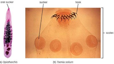
Platyhelminthes: Includes flukes (trematodes) and tapeworms (cestodes)
Flukes: Nonsegmented, oral sucker
Tapeworms: Segmented, scolex with suckers/hooks, proglottids with reproductive structures
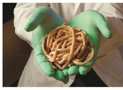
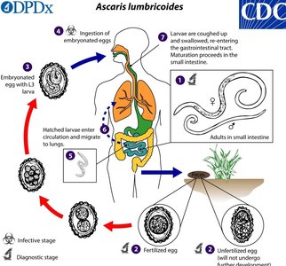
Ascaris lumbricoides
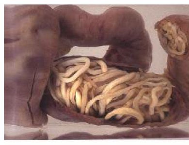
Ascaris lumbricoides is a roundworm that does not attach to the intestinal wall but migrates through tissues, causing inflammation and potential blockages. Its life cycle involves both larval and adult stages within humans, with eggs released in feces and transmitted via contaminated food or objects.
Larvae migrate through intestines, lungs, and pharynx
Adult worms mature in intestines; females produce up to 200,000 eggs/day
Heavy infections can impede development and cause blockages
Ascaris Life Cycle, Prevention, and Treatment
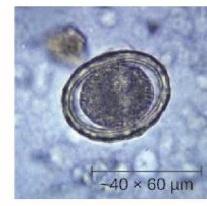
The life cycle of Ascaris lumbricoides involves ingestion of embryonated eggs, migration of larvae through tissues, and maturation in the intestines. Prevention includes handwashing and avoiding contaminated soil. Diagnosis is made by identifying eggs in stool samples, and treatment involves anthelminthic drugs.
Prevention: Hygiene, food safety
Diagnosis: Stool sample analysis
Treatment: Anthelminthic drugs
Fungi
General Characteristics
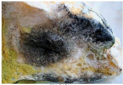
Fungi are heterotrophic organisms, obtaining nutrients from organic matter. Most are saprozoic, living off dead plants and animals, though some are parasitic. Fungal infections (mycoses) are often opportunistic, affecting individuals with weakened immune systems.
Heterotrophic: Require organic nutrients
Saprozoic: Decompose dead matter
Opportunistic pathogens: Cause disease in immunocompromised hosts
Microscopic Fungi: Yeasts and Molds
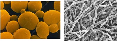
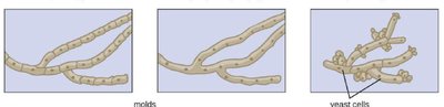
Microscopic fungi exist as yeasts (unicellular, round/ovoid, reproduce asexually) or molds (multicellular, filamentous hyphae forming a mycelium). Some fungi exhibit dimorphism, switching between yeast and mold forms depending on environmental conditions.
Yeast: Unicellular, asexual reproduction
Mold: Multicellular, hyphae and mycelium
Dimorphism: Ability to exist in two forms
Dimorphism and Pathogenic Fungi
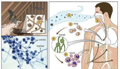
Dimorphic fungi can exist as molds in the environment and as yeasts within host organisms. Histoplasma capsulatum is a dimorphic fungus that causes histoplasmosis, a chronic lung disease. It grows as mycelium in soil and transforms into yeast form in the lungs after inhalation of spores.
Environmental form: Mold/mycelium, cooler temperatures
Host-associated form: Yeast, body temperature
Histoplasma capsulatum: Found in soil, associated with bird/bat droppings
Infection: Inhalation of conidia, pulmonary disease, possible systemic spread

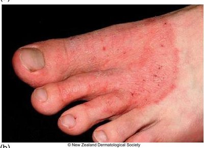
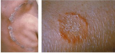
Clinical Focus: Ringworm
Ringworm is a common fungal infection, especially among children and athletes. Despite its name, it is not caused by a worm but by fungi such as Trichophyton rubrum. Diagnosis is confirmed by the presence of cell walls in the specimen, and treatment involves antifungal creams.
Transmission: Close contact, contaminated objects
Diagnosis: Microscopic examination for fungal cell walls
Treatment: Antifungal creams
Causative agent: Trichophyton rubrum
Summary Table: Major Eukaryotic Microorganisms
Group | Key Features | Examples | Diseases |
|---|---|---|---|
Protozoa | Unicellular, motile, parasitic, cyst formation | Giardia, Entamoeba, Plasmodium | Malaria, amoebiasis, giardiasis |
Helminths | Multicellular, organ systems, eggs/larvae | Ascaris lumbricoides, Taenia solium, Opisthorchis | Ascariasis, tapeworm infection, fluke infection |
Fungi | Heterotrophic, saprozoic, yeast/mold, dimorphism | Histoplasma capsulatum, Trichophyton rubrum | Histoplasmosis, ringworm, candidiasis |
Additional info: Academic context was added to clarify classification, life cycles, and clinical relevance of each group. The summary table was inferred for completeness and exam preparation.
