 Back
Eukaryotic vs Prokaryotic Cells: Structure, Morphology, and Specialized Features
Study Guide - Smart Notes
 Tailored notes based on your materials, expanded with key definitions, examples, and context.
Topic 3: Eukaryotic vs Prokaryotic Cells
Overview of Prokaryotic and Eukaryotic Cells
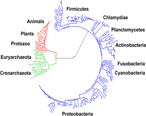
Cells are the fundamental units of life, and they are classified into two main types: prokaryotic and eukaryotic cells. Understanding their differences is essential for microbiology, as it underpins the classification, physiology, and pathogenicity of microorganisms.
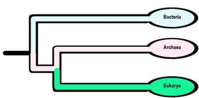
Prokaryotes: Organisms whose cells lack a true nucleus and membrane-bound organelles. Includes Bacteria and Archaea.
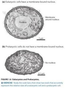
Eukaryotes: Organisms whose cells possess a true nucleus and various membrane-bound organelles. Includes animals, plants, fungi, and protists.
Key Differences:
Feature | Prokaryotes | Eukaryotes |
|---|---|---|
Nucleus | Absent | Present |
Chromosomes | Single, circular, not in a membrane | Multiple, linear, in nuclear membrane |
Histones | Absent | Present |
Organelles | Absent | Present |
Cell Wall | Peptidoglycan (Bacteria), Pseudomurein (Archaea) | Polysaccharide (plants, fungi) |
Division | Binary fission | Mitosis |
Features of Bacterial Cells
Bacteria are the most abundant and diverse organisms on Earth. Their cellular structure is adapted for survival in a wide range of environments.
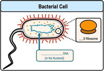
DNA: Circular, located in the nucleoid region.
Ribosomes: 70S type, smaller than eukaryotic ribosomes.
Cell Division: Binary fission.
Size, Shape, and Arrangement of Bacterial Cells
Bacterial morphology is defined by cell size, shape, and arrangement. These characteristics are important for identification and classification.
Size: 0.2–2.0 μm in diameter, 2–8 μm in length.
Shape: Most bacteria are monomorphic (single shape), but some are pleomorphic (variable shapes).
Major Shapes:
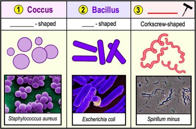
Coccus: Spherical
Bacillus: Rod-shaped
Spiral: Includes vibrio (comma-shaped), spirillum (rigid spiral), and spirochete (flexible spiral)
Others (rare): Star-shaped, rectangular
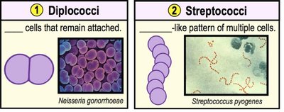
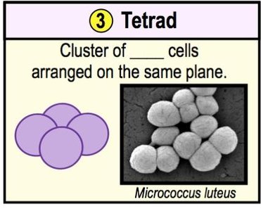
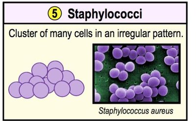
Cell Arrangements:
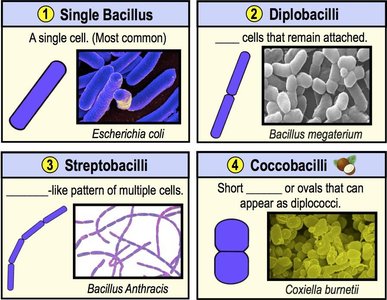
Pairs: Diplococci, diplobacilli
Chains: Streptococci, streptobacilli
Clusters: Staphylococci
Groups of four: Tetrads
Bacterial External Structures
Glycocalyx: Capsules and Slime Layers
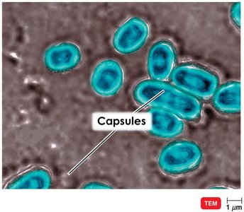
The glycocalyx is a viscous, gelatinous layer external to the cell wall, composed of polysaccharide and/or polypeptide. It enhances bacterial survival and pathogenicity.
Capsule: Neatly organized, firmly attached; protects against phagocytosis and contributes to virulence.
Slime Layer: Unorganized, loosely attached; aids in surface adhesion and biofilm formation.
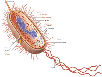
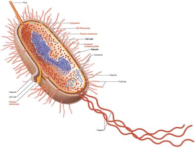
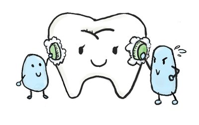
Example: Streptococcus pneumoniae (pneumonia) and Streptococcus mutans (dental caries) use capsules as virulence factors.
Biofilms: Communities of bacteria encased in a polysaccharide matrix attached to surfaces, important in chronic infections and resistance to antimicrobials.
Flagella
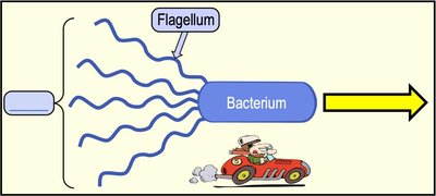
Flagella are long, filamentous appendages that provide motility to bacteria. They are composed of the protein flagellin and are anchored in the cell envelope.
Structure: Three parts—filament, hook, basal body.
Function: Motility (run and tumble), taxis (movement toward/away from stimuli), antigenic properties (H antigens).
Flagellar Arrangements:
Monotrichous: Single flagellum
Lophotrichous: Tuft at one end
Amphitrichous: One or more at both ends
Peritrichous: Distributed over entire cell
Chemotaxis: Movement in response to chemical gradients; bacteria lengthen runs toward attractants and increase tumbles when moving away.
Fimbriae and Pili
Fimbriae and pili are non-motile, proteinaceous projections from the cell surface, important for adhesion and genetic exchange.
Fimbriae: Short, numerous; mediate attachment to surfaces and biofilm formation.
Pili: Longer, fewer; involved in DNA transfer (conjugation pili/sex pili).
Bacterial Cell Wall
Structure and Function
The bacterial cell wall provides structural support, maintains cell shape, and protects against osmotic lysis. Its composition is a key determinant in bacterial classification and antibiotic susceptibility.
Main component: Peptidoglycan—a polymer of N-acetylglucosamine (NAG) and N-acetylmuramic acid (NAM) cross-linked by peptides.
Types of Bacterial Cell Walls
Feature | Gram-Positive | Gram-Negative |
|---|---|---|
Peptidoglycan | Thick (multiple layers) | Thin (single layer) |
Teichoic acids | Present | Absent |
Outer membrane | Absent | Present (contains LPS) |
Susceptibility to penicillin | High | Low |
Lysozyme sensitivity | High | Low |
Endotoxins | Absent | Present (Lipid A) |
Gram Stain Mechanism:
Gram-positive: Alcohol dehydrates peptidoglycan, trapping crystal violet-iodine complex (purple).
Gram-negative: Alcohol dissolves outer membrane, crystal violet-iodine complex washes out, safranin counterstains (pink).
Atypical Cell Walls
Acid-fast bacteria: Mycobacterium and Nocardia; contain mycolic acid (waxy lipid) in cell wall; resist decolorization by acid-alcohol.
Mycoplasmas: Lack cell walls; have sterols in plasma membrane.
Bacterial Plasma Membrane
Structure and Function
The plasma membrane is a phospholipid bilayer with embedded proteins, functioning as a selective barrier and site for metabolic activities.
Fluid mosaic model: Proteins and lipids move laterally; membrane is dynamic and self-sealing.
Functions: Selective permeability, ATP production, photosynthetic pigments (in some bacteria).
Transport Mechanisms
Passive transport: No energy required; includes simple diffusion, facilitated diffusion, and osmosis.
Active transport: Requires energy (ATP); moves substances against concentration gradient (e.g., Na+/K+ pump).
Osmosis: Movement of water across a selectively permeable membrane. Cells in hypotonic solutions gain water; in hypertonic solutions, lose water.
Prokaryotic Internal Structures
Nucleoid and Plasmids
Nucleoid: Region containing the bacterial chromosome (circular DNA).
Plasmids: Small, extrachromosomal DNA molecules; often carry antibiotic resistance or toxin genes.
Ribosomes
70S ribosomes: Sites of protein synthesis; composed of 50S and 30S subunits.
Inclusions
Storage granules: Polysaccharide, lipid, sulfur, phosphate reserves.
Gas vacuoles: Provide buoyancy.
Magnetosomes: Contain iron oxide; orient cells in magnetic fields.
Endospores
Endospores: Dormant, highly resistant structures formed by Bacillus and Clostridium species; survive extreme conditions.
Sporulation: Formation of endospores.
Germination: Return to vegetative state.
Eukaryotic Cell Structures
General Features
Eukaryotic cells are structurally more complex than prokaryotic cells, containing a variety of organelles that compartmentalize cellular functions.
Nucleus: Double-membrane structure containing DNA complexed with histones (chromatin).
Endoplasmic Reticulum (ER): Rough ER (with ribosomes) synthesizes proteins; smooth ER synthesizes lipids and hormones.
Golgi Complex: Modifies, sorts, and packages proteins for secretion or delivery to other organelles.
Lysosomes: Contain digestive enzymes for intracellular digestion.
Vacuoles: Storage and structural support (especially in plants).
Mitochondria: Sites of ATP production via cellular respiration.
Chloroplasts: Sites of photosynthesis in plants and algae.
Cytoskeleton: Provides shape, support, and facilitates movement.
Ribosomes: 80S in cytoplasm and on ER; 70S in mitochondria and chloroplasts.
Cell Wall and Plasma Membrane
Cell Wall: Present in plants (cellulose), fungi (chitin), and some protists; absent in animals.
Plasma Membrane: Phospholipid bilayer with sterols; involved in selective transport and cell signaling.
Specialized Structures
Flagella and Cilia: Used for locomotion or moving substances along the cell surface; flagella are longer and fewer, cilia are shorter and more numerous.
Endocytosis: Uptake of materials via vesicle formation; includes phagocytosis (solid particles) and pinocytosis (liquids).
