 Back
Lab 2: Examination of Living Microorganisms: Microscopy Techniques and Motility
Study Guide - Smart Notes
 Tailored notes based on your materials, expanded with key definitions, examples, and context.
Examination of Living Microorganisms
Introduction
This study guide covers the preparation and observation of living microorganisms using wet-mount and hanging-drop techniques. It explains how to distinguish true motility from Brownian movement and compares different microscopy methods for visualizing live cells. These skills are foundational for microbiology students learning to observe and interpret microbial life.
Microscopy Techniques for Observing Microorganisms
Brightfield Microscopy
Brightfield microscopy is the most common method for observing stained or naturally pigmented specimens. In this technique, the specimen appears dark against a bright background. However, unstained microorganisms are often difficult to see due to low contrast with their aqueous environment.
Key Point: Brightfield is best for stained or pigmented samples; live, unstained cells are hard to distinguish.
Limitation: Low contrast between cells and background makes visualization of live, transparent organisms challenging.
Improving Contrast: Darkfield and Phase-Contrast Microscopy
To observe live, unstained microorganisms, specialized microscopy techniques are used to enhance contrast without staining.
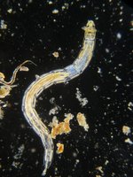
Darkfield Microscopy
Principle: Uses a special condenser with an opaque disk to block direct light; only light scattered by the specimen enters the objective lens, making the specimen appear bright against a dark background.
Applications: Ideal for visualizing organisms that cannot be stained or are distorted by staining, such as Treponema pallidum (syphilis spirochete).
Phase-Contrast Microscopy
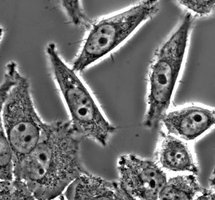
Principle: Converts small differences in refractive index within the specimen into differences in brightness, allowing visualization of internal structures in live cells without staining.
Mechanism: Collects direct, reflected, refracted, and diffracted light to generate a more three-dimensional image.
Advantage: Reveals internal details and motility in living cells.
Resolution in Microscopy
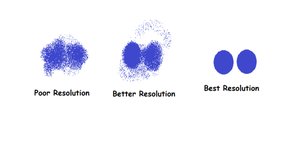
Resolution is the ability of a microscope to distinguish two points as separate entities. Higher resolution allows for clearer and more detailed images of microorganisms.
Poor Resolution: Two points appear merged.
Better Resolution: Two points are distinguishable but not sharply separated.
Best Resolution: Two points are clearly separated.
Light Interactions: Reflection, Refraction, and Diffraction
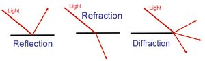
Microscopy relies on the interaction of light with specimens. Three key phenomena are:
Reflection: Light bounces off the surface of a specimen.
Refraction: Light bends as it passes through materials of different densities.
Diffraction: Light spreads as it passes through small openings or edges, contributing to image formation and resolution.
Distinguishing Motility in Microorganisms
Brownian Movement vs. True Motility
It is important to differentiate between random motion caused by molecular collisions (Brownian movement) and active movement by microorganisms (true motility).
Brownian Movement: Caused by water molecules striking particles; all particles vibrate at the same rate and maintain relative positions. Movement is random and non-directional.
True Motility: Organisms actively move from one position to another, often in a directed manner. Cells may spin, roll, or swim independently.
Note: Fluid currents in a wet mount can move all particles in one direction; this is not true motility.
Slide Preparation Techniques
Wet-Mount Technique
The wet-mount technique is a simple method for observing live microorganisms in a drop of liquid.
Stir or shake the infusion gently to suspend organisms.
Transfer a small drop to a slide using a Pasteur pipette or sterilized inoculating loop.
Place a coverslip on the drop, handling it by the edges.
Press gently to spread the liquid.
Observe under 10×, then 40×, and finally 100× (oil immersion) objectives, adjusting light as needed.
Record observations, noting size, shape, color, and movement.
Hanging-Drop Technique
The hanging-drop method allows for better observation of motility and three-dimensional structure by suspending a drop of liquid from a coverslip over a depression slide.
Advantages: Greater depth, reduced evaporation, less interference from surface tension and fluid currents. Ideal for observing motility in bacteria such as Bacillus.
Procedure:
Apply petroleum jelly to the edges of a coverslip.
Place a drop of sample in the center of the coverslip.
Invert a depression slide over the coverslip and flip so the drop hangs in the well.
Observe at low and then higher magnification, recording observations.
Note: Do not use the 100× oil immersion lens with hanging-drop slides.
Laboratory Materials and Safety
Supplies and Cultures
Slides, coverslips, hanging-drop (depression) slides
Petroleum jelly, toothpick, Pasteur pipettes, inoculating loop, alcohol, Gram's iodine, lens paper
Cultures: hay infusion (light and dark), peppercorn infusion, Bacillus broth culture
Cleanup and Disposal
Remove slides from microscope, lower stage, rotate to 4×, wrap cord, and store properly.
Discard slides and coverslips in designated containers; disinfect as required.
Recording and Interpreting Results
Notebook Requirements
Draw and label two wet-mount observations from different infusions, noting color, movement, and magnification.
Draw and label two hanging-drop observations: one from an infusion, one from Bacillus culture, both at 400×.
Write conclusions: compare results to expectations, describe how motility was distinguished, and note which infusion had the most organisms.
Summary Table: Comparison of Microscopy Techniques
Technique | Contrast Mechanism | Best For | Limitations |
|---|---|---|---|
Brightfield | Absorption of light by stained or pigmented cells | Stained or naturally pigmented specimens | Poor for live, unstained cells |
Darkfield | Scattered light from specimen | Unstained, delicate, or thin organisms | Limited internal detail |
Phase-Contrast | Differences in refractive index | Live, unstained cells; internal structures | Requires specialized optics |
Additional info:
Hay infusions incubated in light are likely to have more photosynthetic microorganisms (e.g., algae) compared to those incubated in the dark.
Understanding the difference between Brownian movement and true motility is essential for interpreting microbial behavior in environmental and clinical samples.
