 Back
BackFoundations of Microbiology and Microbial Genetics
Study Guide - Smart Notes
 Tailored notes based on your materials, expanded with key definitions, examples, and context.
Introduction to Microbiology
Definition and Scope
Microbiology is the branch of science that deals with organisms too small to be seen by the naked eye, known as microorganisms or microbes. These include bacteria, viruses, fungi, protozoa, and algae. Microbiology explores their structure, function, genetics, and roles in health, disease, and the environment.
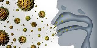
Microorganisms are found everywhere: in the air, water, food, on our skin, and inside our bodies.
Many microbes are beneficial, aiding in processes such as vitamin production, waste decomposition, and atmospheric maintenance.
Some microbes are pathogenic and can cause diseases.
Presence of Microorganisms in Daily Life
Air: Microbes are present in the air we breathe.
Water: Drinking water can contain various microorganisms.
Food: Microbes are found on and in food items.
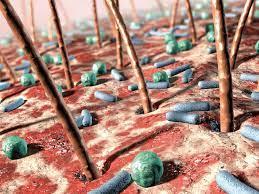
Skin: The skin hosts a diverse microbial community.
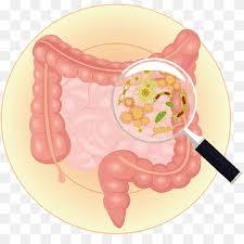
Body: The human body, especially the gut, contains trillions of microbes essential for health.
Microbial Genetics
Overview and Importance
Microbial genetics is the study of how microorganisms inherit traits, how their genetic material is organized, replicated, and expressed, and how genetic information is transferred between cells. This field is fundamental for understanding microbial physiology, evolution, and biotechnology applications.
Genetics is the science of DNA and its role in heritable cellular features, activities, and variations.
Key molecules: DNA, RNA, and proteins.
Gene transfer, expression, and regulation are central topics.
History of DNA Discovery
Frederick Griffith (1928): Demonstrated transformation in bacteria, suggesting genetic information could be transferred between cells.
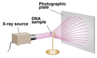
Rosalind Franklin (1952): Produced X-ray diffraction images of DNA, revealing its helical structure.
Watson and Crick (1953): Described the double helix structure of DNA, using Franklin's data.
Maurice Wilkins: Contributed to the elucidation of DNA structure; shared the Nobel Prize with Watson and Crick in 1962.
Chemical Structure of DNA
Nucleotides and DNA Structure
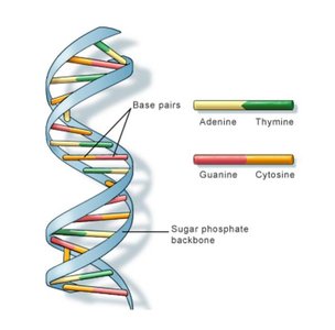
DNA (Deoxyribonucleic Acid) is a polymer composed of repeating units called nucleotides. Each nucleotide consists of a deoxyribose sugar, a phosphate group, and a nitrogenous base. DNA is structured as a double helix with two antiparallel strands held together by hydrogen bonds between complementary bases.
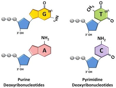
Nucleotides: Building blocks of DNA, each containing:
A five-carbon sugar (deoxyribose)
A phosphate group
A nitrogenous base (Adenine, Thymine, Cytosine, Guanine)
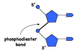
Phosphodiester bonds link nucleotides in a strand (5' phosphate to 3' hydroxyl).
Base pairing: Adenine (A) pairs with Thymine (T); Cytosine (C) pairs with Guanine (G).
Antiparallel strands: One strand runs 5' to 3', the other 3' to 5'.
Base Pairing and Stability
Hydrogen bonds stabilize the double helix:
A-T pairs form two hydrogen bonds.
G-C pairs form three hydrogen bonds (more stable).
Complementary base pairing ensures accurate DNA replication and transcription.
DNA Supercoiling
Supercoiling refers to the over- or under-winding of the DNA double helix. In bacteria, the enzyme DNA gyrase (a type II topoisomerase) introduces negative supercoils, aiding in the compact packaging of DNA within the cell.
Supercoiling is essential for fitting large DNA molecules into small cellular spaces.
Topoisomerases regulate DNA supercoiling during replication and transcription.
Central Dogma of Molecular Biology
Flow of Genetic Information
The central dogma describes the flow of genetic information within a biological system:
Replication: DNA makes an identical copy of itself.
Transcription: DNA is used as a template to synthesize RNA.
Translation: Messenger RNA (mRNA) directs protein synthesis.
DNA Replication
Mechanism and Enzymes
DNA replication is the process by which a cell duplicates its DNA before cell division. It is semi-conservative, meaning each new DNA molecule consists of one old (parental) strand and one newly synthesized strand.
Initiation: Replication begins at specific sites called origins of replication. DNA helicase unwinds the double helix, and DNA primase synthesizes short RNA primers to initiate synthesis.
Elongation: DNA polymerase adds nucleotides to the 3' end of the primer, synthesizing the new strand in a 5' to 3' direction. The leading strand is synthesized continuously, while the lagging strand is synthesized in short fragments (Okazaki fragments).
Termination: Replication ends when the entire DNA molecule has been copied. On the lagging strand, RNA primers are removed by RNase H, and DNA ligase joins Okazaki fragments to form a continuous strand.
Feature | Leading Strand | Lagging Strand |
|---|---|---|
Synthesis Direction | 5' to 3' (continuous) | 5' to 3' (discontinuous) |
Primer Requirement | One primer | Multiple primers |
Fragments | None | Okazaki fragments |
Enzyme for Joining | Not required | DNA ligase |
Key enzymes: DNA helicase, DNA primase, DNA polymerase, RNase H, DNA ligase, DNA gyrase (topoisomerase II).
Equation for DNA synthesis:
Additional info: dNTP = deoxynucleoside triphosphate; PPi = pyrophosphate.
