 Back
Foundations of Microbiology: Key Concepts, Methods, and Microbial Diversity
Study Guide - Smart Notes
 Tailored notes based on your materials, expanded with key definitions, examples, and context.
Branches of Microbiology
Overview of Microbiology Branches
Microbiology is a diverse field with several specialized branches, each focusing on different aspects of microorganisms and their interactions with humans, animals, plants, and the environment.
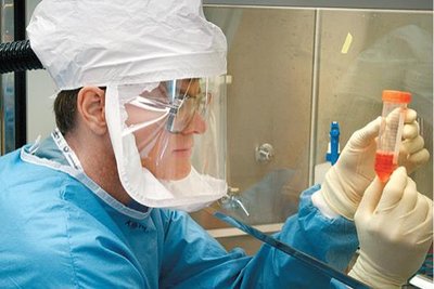
Medical Microbiology: Studies microbes that cause diseases in humans and animals, focusing on diagnosis, treatment, and prevention.
Agricultural Microbiology: Examines the relationships between microbes and crops or farm animals, including soil fertility and plant diseases.
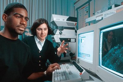
Immunology: Investigates the immune system's response to microbial infections, including the production of antibodies and immune cells.
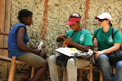
Public Health Microbiology and Epidemiology: Monitors and controls the spread of infectious diseases in communities.
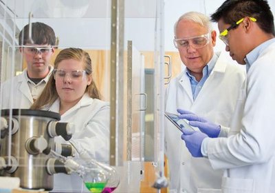
Industrial Microbiology: Ensures the safety of food and water, and applies biotechnology for the production of antibiotics, enzymes, and other products.
Environmental Microbiology: Studies the effects of microbes on diverse habitats such as soil, water, and extreme environments (geomicrobiology, astrobiology).
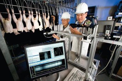
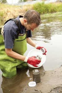
Examples of Branches in Action
Medical Microbiology: Investigating outbreaks caused by Salmonella enterica in contaminated food.
Agricultural Microbiology: Addressing fungal fruit rot in crops.
Public Health Microbiology: Managing community-wide outbreaks through surveillance and intervention.
Immunology: Using blood tests to detect immune responses to pathogens.
Industrial Microbiology: Producing fermented foods like cheese and wine.
Environmental Microbiology: Assessing soil microbial balance for optimal plant growth.
Golden Age Microbiologists and Their Contributions
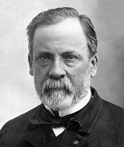
Louis Pasteur
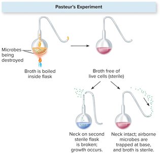
Louis Pasteur made foundational contributions to microbiology, including disproving spontaneous generation, inventing pasteurization, and developing the germ theory of disease. He also created the first rabies vaccine.
Pasteurization: Heating liquids to kill harmful microbes without altering the liquid's properties.
Germ Theory: Proposed that specific microorganisms cause specific diseases.
Disproving Spontaneous Generation: Demonstrated that microbes do not arise spontaneously but from other microbes.
Other Key Scientists
Robert Hooke: First to observe microbes using a microscope.
Antonie van Leeuwenhoek: Developed simple microscopes and described "animalcules" (microorganisms) from water and dental scrapings.
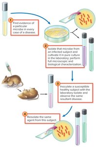
Robert Koch: Established Koch's postulates, linking specific microbes to specific diseases.
John Needham: Attempted to disprove spontaneous generation but failed due to incomplete sterilization.
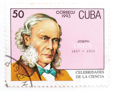
Joseph Lister: Pioneered antiseptic techniques in surgery, including handwashing and using antiseptic chemicals.
John Tyndall: Showed that some microbes are highly heat-resistant, requiring more than boiling to destroy.
Oliver Wendell Holmes & Ignaz Semmelweis: Demonstrated the importance of hand hygiene in preventing hospital-acquired infections.

Microbial Nomenclature
Binomial System of Nomenclature
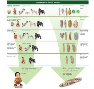
The binomial system, established by Carl von Linne (Linnaeus), assigns each organism a two-part scientific name: the genus (capitalized) and the species (lowercase), both italicized (e.g., Staphylococcus aureus).
Genus: Group of closely related species.
Species: Specific organism within the genus.
Microscopy in Microbiology
Types of Light and Electron Microscopes
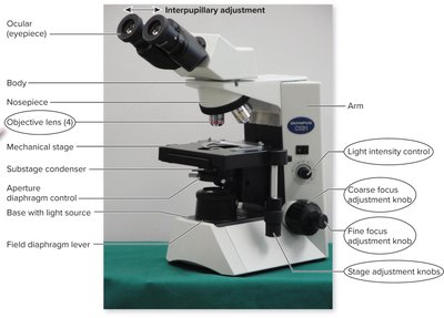
Microscopes are essential tools for observing microorganisms. Different types provide varying levels of detail and are suited for specific applications.
Light Microscopes:
Bright-field: Most common; used for stained specimens.
Dark-field: Visualizes live, unstained cells.
Phase-contrast: Highlights intracellular structures.
Interference (DIC): Produces 3D-like images.
Fluorescent: Uses fluorescent dyes to visualize specific structures.
Confocal: Provides detailed, layered images using lasers.
Electron Microscopes:
Transmission (TEM): Reveals internal cell structures and viruses.
Scanning (SEM): Produces dramatic 3D images of surfaces.
Staining Techniques in Light Microscopy
Staining enhances contrast in microscopic specimens, allowing for better visualization and differentiation of microbial structures.
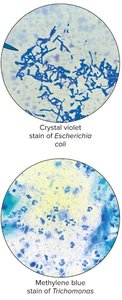
Simple Stains: Use a single dye to color cells, revealing shape and arrangement.
Differential Stains: Use multiple dyes to distinguish between cell types or structures.
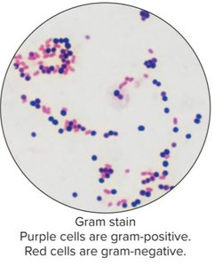
Gram Stain: Differentiates bacteria into Gram-positive (purple) and Gram-negative (red) based on cell wall structure.
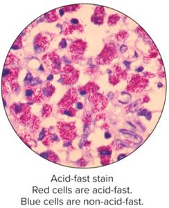
Acid-fast Stain: Identifies acid-fast bacteria (e.g., Mycobacterium).
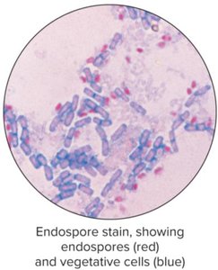
Endospore Stain: Highlights bacterial endospores (red) and vegetative cells (blue).
Microbial Genetics
DNA Replication, Transcription, and Translation
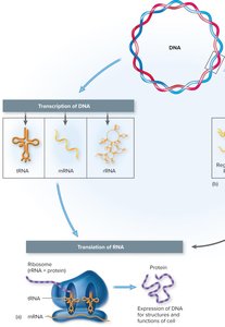
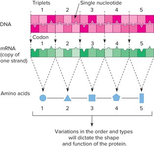
Genetic information in microorganisms is stored in DNA and expressed through the processes of replication, transcription, and translation.
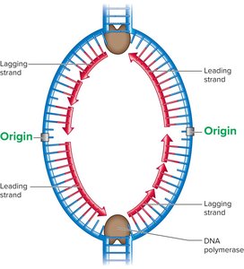
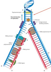
DNA Replication: The process by which DNA makes an identical copy of itself before cell division.
Enzymes involved: helicase (unwinds DNA), primase (synthesizes RNA primer), DNA polymerase III (adds nucleotides), DNA ligase (seals gaps).
Replication is semi-conservative: each new DNA molecule contains one parental and one new strand.
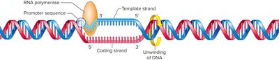
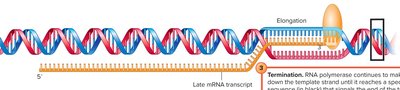
Transcription: DNA is transcribed into messenger RNA (mRNA) by RNA polymerase.
Translation: mRNA is decoded by ribosomes to synthesize proteins, following the genetic code (codons specify amino acids).
Horizontal Gene Transfer in Bacteria
Horizontal gene transfer (HGT) allows bacteria to acquire new genetic traits from other organisms, contributing to genetic diversity and adaptation.
Mode | Factors Involved | Direct/Indirect | Examples of Products |
|---|---|---|---|
Conjugation | Donor cell with pilus; fertility plasmid; both cells alive; DNA bridge forms | Direct | Drug resistance, toxin production, adherence molecules |
Transformation | Free donor DNA; live, competent recipient | Indirect | Capsule formation, genetic engineering products |
Transduction | Donor lysed; bacteriophage transfers DNA; recipient of same species | Indirect | Toxins, drug resistance |
Transposons | "Jumping genes" move within or between DNA molecules | Indirect | Genetic variability, antibiotic resistance |
Mutations and Their Effects
Mutations are changes in the DNA sequence that can affect microbial traits.
Spontaneous Mutations: Occur naturally due to errors in DNA replication.
Induced Mutations: Result from exposure to mutagens (chemicals or radiation).
Mutagen | Effect |
|---|---|
Nitrous acid, bisulfite | Remove amino groups from bases |
Ethidium bromide | Inserts between base pairs |
Acridine dyes | Cause frameshifts |
Nitrogen base analogs | Compete with natural bases |
Ionizing radiation | Breaks DNA strands |
Ultraviolet | Causes pyrimidine dimers |
Point Mutations: Affect a single base (addition, deletion, or substitution).
Lethal Mutation: Causes cell death or dysfunction.
Neutral Mutation: No significant effect on cell function.
Missense Mutation: Changes one amino acid in a protein.
Nonsense Mutation: Converts a codon to a stop codon, truncating the protein.
Silent Mutation: Alters DNA but not the amino acid sequence.
Frameshift Mutation: Insertion or deletion (not in multiples of three) shifts the reading frame, usually rendering the protein nonfunctional.
Cellular Organization of Microorganisms
Prokaryotic Cell Structure
Bacterial cells have specialized structures that contribute to their survival, pathogenicity, and classification.
External Structures: Flagella (motility), fimbriae and pili (attachment and DNA transfer).
Surface Coatings: Capsules and slime layers protect against desiccation and immune attack.
Cell Envelope: Includes the cell wall (Gram-positive or Gram-negative) and cell membrane.
Internal Structures: Cytoplasm, ribosomes, nucleoid (DNA), plasmids (extra-chromosomal DNA), endospores (dormant, resistant forms).
Gram-Positive vs. Gram-Negative Cell Walls
Gram-Positive: Thick peptidoglycan layer, teichoic acids, stains purple.
Gram-Negative: Thin peptidoglycan, outer membrane with lipopolysaccharide (LPS), stains red.
Roles in Pathogenicity
Capsules: Prevent phagocytosis by host immune cells.
Fimbriae: Aid in attachment to host tissues.
Flagella: Enable motility and colonization.
Plasmids: Carry genes for antibiotic resistance and toxins.
Endospores: Allow survival in harsh conditions.
Three Domains of Life
Bacteria, Archaea, and Eukarya
All life is classified into three domains based on cellular organization and genetic characteristics.
Characteristic | Bacteria | Archaea | Eukarya |
|---|---|---|---|
Cell type | Bacterial | Archaeal | Eukaryotic |
Chromosomes | Single/few, circular | Single, circular | Several, linear |
Ribosomes | 70S | 70S (structure similar to 80S) | 80S |
Flagella | Hook, rings, hollow filament | Solid, fimbrial-like | 9+2 microtubule arrangement |
Peptidoglycan in cell wall | Yes | No | No |
Membrane lipids | Ester-linked fatty acids | Ether-linked branched hydrocarbons | Ester-linked fatty acids |
Sterols in membrane | No (some exceptions) | No | Yes |
Environments of Archaea
Archaea are often found in extreme environments, such as hot springs, salt lakes, and deep-sea vents, but also in more common habitats.
Eukaryotic Microorganisms
Cellular Organization, Cell Wall Composition, and Reproduction
Group | Complexity | Cell Wall | Nutritional Type | Motility | Key Structures |
|---|---|---|---|---|---|
Protozoa | Unicellular | None | Heterotrophic/Autotrophic | Flagella, cilia, pseudopodia | Cysts |
Fungi | Uni-/Multicellular | Chitin or cellulose | Heterotrophic | Flagella (gametes) | Hyphae, spores |
Algae | Uni-/Multicellular | Cellulose | Heterotrophic/Autotrophic | Flagella (gametes) | Chloroplasts |
Helminths | Multicellular (adults) | None | Heterotrophic | Limbs | Ova |
Fungal Reproduction
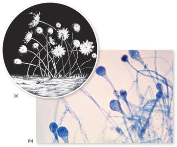
Sporangiospores: Formed inside a sporangium; released when the stalk bursts.
Conidiospores (Conidia): Pinch off from hyphae; not enclosed in a sac.
Helminths
Flatworms: Segmented or unsegmented, thin bodies (e.g., tapeworms, flukes).
Roundworms: Cylindrical, unsegmented bodies (e.g., nematodes).
Helminths can be male, female, or hermaphroditic (both sex organs in one worm).
Acellular Infectious Agents
Viruses
Viruses are noncellular infectious agents observed only with electron microscopy. They consist of genetic material (DNA or RNA) surrounded by a protein coat (capsid), and sometimes an envelope with spikes.
Capsid: Protein shell protecting the genetic material.
Envelope: Lipid membrane derived from host cell, present in some viruses.
Spikes: Glycoproteins for attachment to host cells.
Viral Multiplication Cycle (Animal Viruses)
Adsorption: Virus attaches to host cell receptors.
Penetration: Virus or its genetic material enters the host cell.
Uncoating: Viral capsid is removed, releasing nucleic acid.
Synthesis and Assembly: Viral components are produced and assembled.
Release: New viruses exit the cell by lysis or budding.
Types of Viral Genomes and Replication Strategies
Class | Genome | Strategy | Example Virus | Disease |
|---|---|---|---|---|
1 | dsDNA | dsDNA to mRNA | Herpes simplex | Herpes |
2 | ssDNA | ssDNA to dsDNA to mRNA | Parvovirus B19 | Skin rash |
3 | dsRNA | dsRNA to mRNA | Rotavirus | Gastroenteritis |
4 | (+)ssRNA | (+)ssRNA to (−)ssRNA to mRNA | Poliovirus | Polio |
5 | (−)ssRNA | (−)ssRNA to mRNA | Influenza virus | Influenza |
6 | ssRNA-RT | ssRNA-RT to dsDNA to mRNA | HIV | AIDS |
7 | dsDNA-RT | dsDNA-RT to mRNA | Hepatitis B virus | Hepatitis B |
Persistent and Latent Viral Infections
Persistent Infections: Virus remains in the host cell for extended periods, sometimes for life.
Latency: Virus remains dormant and can reactivate (e.g., herpes simplex, varicella-zoster).
Provirus: Viral DNA integrated into host genome.
Prions and Viroids
Prions: Infectious proteins causing neurodegenerative diseases in animals (e.g., mad cow disease).
Viroids: Infectious naked RNA molecules that infect plants.
Both are noncellular and lack the structures of true viruses.
