 Back
Foundations of Microbiology: Taxonomy, Microscopy, and Bacterial Cell Structure
Study Guide - Smart Notes
 Tailored notes based on your materials, expanded with key definitions, examples, and context.
Introduction to Microbiology
Overview of Microbiology
Microbiology is the study of microscopic organisms, including bacteria, archaea, fungi, protozoa, viruses, and prions. These organisms play essential roles in ecosystems, human health, and disease. Understanding their classification, structure, and physiology is foundational for medical and clinical applications.
Taxonomy and Classification of Microorganisms
Principles of Taxonomy
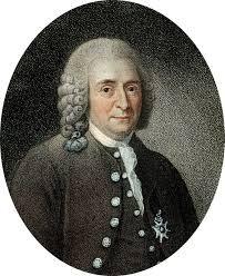
Taxonomy is the science of classifying organisms based on shared characteristics. Early classification relied on morphology (shape, size, arrangement) and physiology. Carl Linnaeus, known as the "Father of Taxonomy," established the binomial nomenclature system, which is still used today.
Taxonomic Hierarchy: Domain, Kingdom, Phylum, Class, Order, Family, Genus, Species
Domains: Bacteria (unicellular prokaryotes), Archaea (unicellular prokaryotes, often extremophiles), Eukarya (unicellular and multicellular eukaryotes)
Prokaryotic species: Defined by physical characteristics and at least 70% DNA similarity, or 97% identical 16S rRNA sequence
Binomial nomenclature: Genus (capitalized) + species (lowercase), italicized (e.g., Escherichia coli)
Strains: Genetic variants within a species, often denoted by numbers/letters (e.g., E. coli K-12)

Types of Microorganisms
Microorganisms are classified into several groups based on cell structure and genetic relationships:
Bacteria: Prokaryotic, diverse morphologies
Archaea: Prokaryotic, often extremophiles, no known pathogens
Fungi: Eukaryotic, includes yeasts and molds
Protozoa: Eukaryotic, often motile
Viruses: Acellular, require host cells for replication
Prions: Infectious proteins, cause neurodegenerative diseases
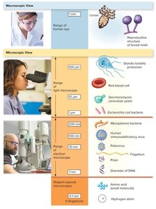
Relative Size of Microorganisms
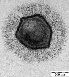
Microorganisms vary greatly in size, from large bacteria visible under light microscopy to viruses and prions requiring electron microscopy.
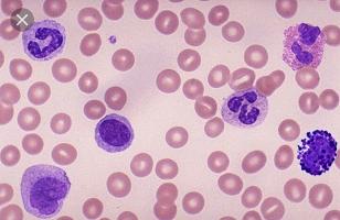
White blood cell: 12–18 μm
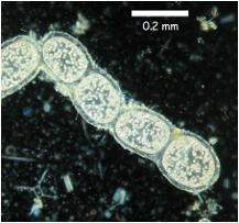
Thiomargarita namibiensis (large bacterium): 100–300 μm
Mimivirus (large virus): 750 nm
Mycoplasma bacteria: ~200 nm
Poliovirus: ~30 nm
Microscopy in Microbiology
Light Microscopy
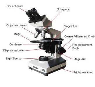
Light microscopy uses visible light to illuminate specimens and is essential for observing cell morphology and arrangement. The compound light microscope is the most common type, using a series of lenses to magnify specimens up to 1,500X with a resolution of about 200 nm.
Objective lenses: 4X, 10X, 40X, 100X (oil immersion)
Ocular lens: Usually 10X
Condenser lens: Focuses light onto the specimen
Iris diaphragm: Adjusts light intensity and contrast
Coarse and fine focus knobs: For rough and precise focusing
Immersion oil: Used with 100X objective to match refractive index of glass, reducing light scatter and improving resolution
Microscopy Techniques
Technique | Image Appearance | Notes |
|---|---|---|
Bright Field | Dark image on bright background | Requires staining; most common |
Dark Field | Light specimen on dark background | Visualizes unstained specimens |
Phase Contrast | Light specimen on dark background | Enhances contrast in unstained cells |
Differential Interference Contrast | 3D-like appearance | Uses polarized light for enhanced contrast |
Electron Microscopy
Electron microscopy uses electron beams for much higher resolution imaging (down to 1 nm). Two main types are:
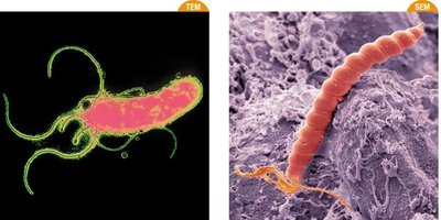
Transmission Electron Microscope (TEM): Electrons pass through specimen, producing 2D images of internal structures. Up to 1,000,000X magnification.
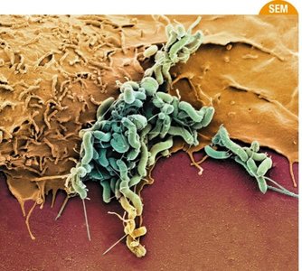
Scanning Electron Microscope (SEM): Electrons scan the surface, producing 3D images of external structures.
Bacterial Morphology and Arrangement
Prokaryotic Cell Shapes
Bacteria exhibit a variety of shapes and arrangements, which are important for identification and classification.
Coccus: Spherical
Bacillus: Rod-shaped
Vibrio: Curved rod
Spirillum: Wave-like or corkscrew
Spirochete: Flexible, corkscrew-shaped
Monomorphic: Single, consistent shape
Pleomorphic: Variable shapes
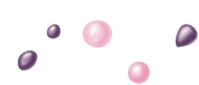
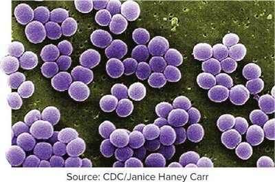
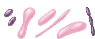
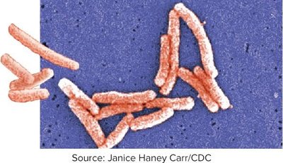
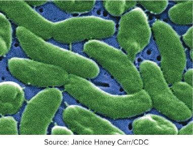
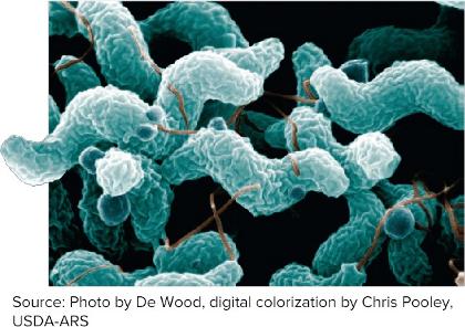
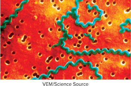
Cell Arrangements
Diplo-: Pairs (e.g., diplococcus, diplobacillus)
Strepto-: Chains (e.g., streptococcus, streptobacillus)
Staphylo-: Clusters (e.g., staphylococcus)
Staining Techniques in Microbiology
Purpose of Staining
Staining increases contrast, making cells and structures more visible under the microscope. Most stains are either basic (positively charged, bind to cell surfaces) or acidic (negatively charged, stain the background).
Basic dyes: Methylene blue, crystal violet, safranin, malachite green
Acidic dyes: Nigrosin, India ink (used for negative staining)
Types of Staining Techniques
Simple Stain: Uses one dye to determine size, shape, and arrangement
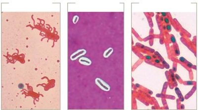
Structural Stains: Highlight specific structures (e.g., flagella, capsules, endospores)
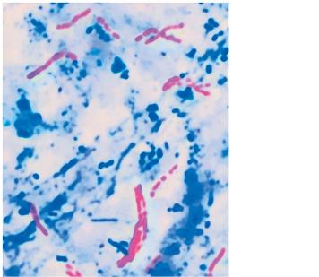
Differential Stains: Distinguish between different types of bacteria (e.g., Gram stain, acid-fast stain)
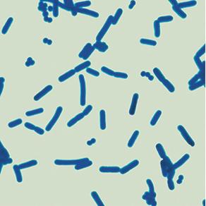
Gram Stain
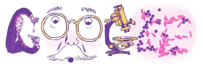
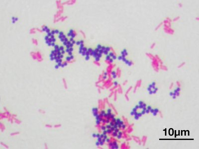
The Gram stain, developed by Hans Christian Gram, is a differential staining technique that distinguishes bacteria based on cell wall structure.
Gram-positive: Thick peptidoglycan layer, stains purple
Gram-negative: Thin peptidoglycan layer, outer membrane, stains pink
Bacterial Cell Structure
Major Components
Plasma membrane: Phospholipid bilayer with proteins; semi-permeable
Cell wall: Provides rigidity and shape; contains peptidoglycan in bacteria
Cytoplasm: Gel-like substance inside the cell
Ribosomes: Sites of protein synthesis
Nucleoid: Region containing the bacterial chromosome
Capsule: Protective outer layer in some bacteria
Fimbriae and flagella: Surface structures for attachment and motility
Cell Wall Structure and Function
The bacterial cell wall is essential for maintaining cell shape, protecting against osmotic lysis, and serving as an antibiotic target. Peptidoglycan is the main structural component.
Gram-positive cell wall: Thick peptidoglycan, teichoic acids
Gram-negative cell wall: Thin peptidoglycan, outer membrane with lipopolysaccharide (LPS), porins
Feature | Gram-Negative | Gram-Positive |
|---|---|---|
Outer membrane | Yes | No |
Lipid A (endotoxin) | Yes | No |
Porins | Yes | No |
Teichoic acids | No | Yes |
Peptidoglycan | Thin (10–20%) | Thick (70–80%) |
Gram stain color | Pink | Purple |
Resistance to drying | No | Yes |
Penicillin susceptibility | Low | High |
Special Cell Wall Types
Acid-fast bacteria: Contain mycolic acid; detected by acid-fast stain (e.g., Mycobacterium)
Cell wall-deficient bacteria: Mycoplasma species lack cell walls, have sterol-rich membranes
L-forms: Bacteria that have lost their cell wall; can persist in harsh conditions
Plasma Membrane Function and Transport
Membrane Structure and Function
The plasma membrane is a phospholipid bilayer with embedded proteins, serving as a selective barrier and site for energy generation (proton motive force).
Transport Mechanisms
Simple diffusion: Passive movement of small, nonpolar molecules down their concentration gradient
Facilitated diffusion: Passive movement via membrane proteins (channels/carriers)
Osmosis: Diffusion of water across the membrane
Active transport: Requires energy (ATP or ion gradients) to move substances against their gradient
Phosphotransferase system: Group translocation; substance is chemically modified during transport (e.g., glucose uptake)
Osmotic Environments
Isotonic: No net water movement
Hypertonic: Water leaves cell, causing plasmolysis
Hypotonic: Water enters cell, risk of lysis if cell wall is damaged
Historical Foundations of Microbiology
Key Figures and Discoveries
Robert Hooke: First compound microscope, coined "cell"
Antonie van Leeuwenhoek: First to observe microbes ("animalcules")
Louis Pasteur: Disproved spontaneous generation, developed pasteurization, proposed Germ Theory of Disease
Robert Koch: Developed Koch's postulates for linking microbes to disease
Hans Christian Gram: Developed Gram stain
Alexander Fleming: Discovered penicillin
Koch's Postulates
The suspected pathogen must be present in all cases of the disease and absent from healthy individuals.
The pathogen must be isolated and grown in pure culture.
The cultured pathogen must cause disease when introduced into a healthy host.
The pathogen must be re-isolated from the experimentally infected host and identified as identical to the original.
Clinical and Public Health Relevance
Pathogens and Infectious Disease
Pathogens: Microbes that cause disease; only a small fraction of microbes are pathogenic
Opportunistic pathogens: Cause disease in weakened hosts
Emerging diseases: New or increasing in incidence (e.g., SARS, MERS, COVID-19)
Antibiotic resistance: Growing concern in clinical microbiology
Case Study Example: Cholera Outbreak
Diagnosis of Vibrio cholerae (Gram-negative, comma-shaped bacillus) in a patient with severe diarrhea is confirmed by microscopy and Gram staining. Treatment includes oral rehydration therapy to replace lost ions and water.
Additional info: This guide covers foundational concepts in microbiology, including taxonomy, microscopy, bacterial structure, and clinical relevance, providing a comprehensive overview for exam preparation.
