 Back
BackFunctional Anatomy of Prokaryotic and Eukaryotic Cells: Microbiology Study Guide
Study Guide - Smart Notes
 Tailored notes based on your materials, expanded with key definitions, examples, and context.
Functional Anatomy of Prokaryotic and Eukaryotic Cells
Overview of Cell Types
This section introduces the fundamental differences between prokaryotic and eukaryotic cells, which are central to understanding microbial structure and function.
Prokaryotes: Characterized by a single circular chromosome not enclosed in a membrane, lack histones and organelles, possess peptidoglycan cell walls (in bacteria), and reproduce by binary fission.
Eukaryotes: Have paired chromosomes within a nuclear membrane, contain histones and organelles, possess polysaccharide cell walls, and divide by mitosis.
Terminology: 'Prokaryote' means 'prenucleus'; 'Eukaryote' means 'true nucleus'.
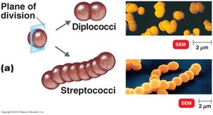
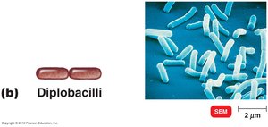
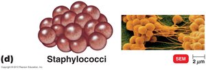
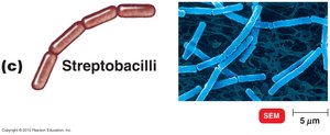
Shapes and Arrangements of Prokaryotic Cells
Bacterial morphology is diverse, with cells exhibiting characteristic shapes and arrangements that aid in identification and classification.
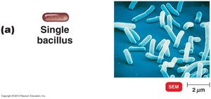
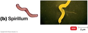
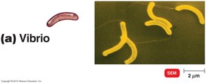
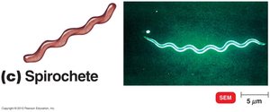
Shapes: Bacillus (rod-shaped), Coccus (spherical), Spiral (spirillum, vibrio, spirochete).
Arrangements: Diplococci (pairs), Streptococci (chains), Staphylococci (clusters), Diplobacilli (pairs), Streptobacilli (chains).
Monomorphic: Most bacteria have a single shape; Pleomorphic: Some can vary in shape.
Examples: Streptococcus forms chains of cocci; Bacillus forms rods.

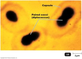

Glycocalyx: Capsule and Slime Layer
The glycocalyx is a protective and adhesive layer outside the cell wall, crucial for bacterial survival and pathogenicity.
Capsule: Neatly organized, prevents phagocytosis.
Slime Layer: Unorganized and loose, aids in attachment.
Extracellular Polysaccharide: Facilitates cell attachment to surfaces.
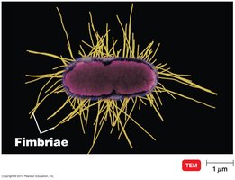
Flagella, Fimbriae, and Pili
These surface structures are essential for motility, attachment, and genetic exchange in prokaryotes.
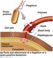
Flagella: Long, whip-like appendages for movement; composed of flagellin, attached via a basal body and hook.
Fimbriae: Short, hair-like structures for attachment to surfaces.
Pili: Longer than fimbriae, facilitate DNA transfer (conjugation).
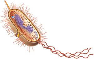
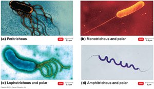
Arrangements of Bacterial Flagella
Bacteria exhibit various flagellar arrangements, which influence their motility and identification.
Peritrichous: Flagella distributed over the entire cell.
Monotrichous: Single flagellum at one pole.
Lophotrichous: Tuft of flagella at one pole.
Amphitrichous: Flagella at both poles.
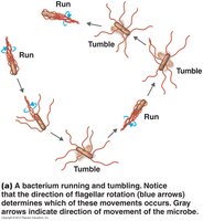

Motility and Chemotaxis
Bacterial motility is achieved by rotating flagella, enabling movement toward or away from stimuli (taxis).
Run and Tumble: Alternating straight movement (run) and random change in direction (tumble).
Flagella Proteins: Serve as H antigens for identification (e.g., E. coli O157:H7).
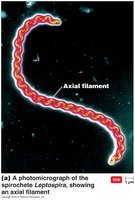
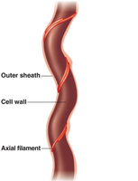
Axial Filaments, Fimbriae, and Pili
Axial filaments are unique to spirochetes, enabling corkscrew motility. Fimbriae and pili are involved in attachment and genetic exchange.
Axial Filaments: Endoflagella anchored at one end, rotation causes cell movement.
Gliding and Twitching Motility: Alternative forms of movement.

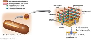
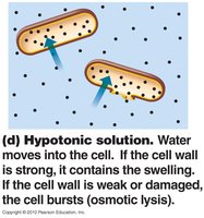
The Cell Wall: Structure and Function
The cell wall is a critical structure for bacterial integrity, preventing osmotic lysis and providing shape.
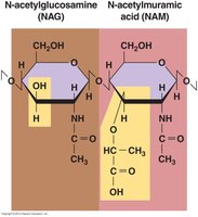
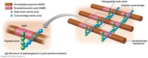
Peptidoglycan: Polymer of N-acetylglucosamine (NAG) and N-acetylmuramic acid (NAM), linked by polypeptides.
Function: Maintains cell shape, protects against osmotic pressure.
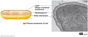
Gram-Positive vs. Gram-Negative Cell Walls
Bacteria are classified based on cell wall structure, which affects staining, susceptibility to antibiotics, and pathogenicity.
Gram-Positive: Thick peptidoglycan, teichoic acids, disrupted by lysozyme, sensitive to penicillin.
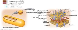
Gram-Negative: Thin peptidoglycan, outer membrane with lipopolysaccharides (LPS), endotoxin (Lipid A), sensitive to tetracycline.


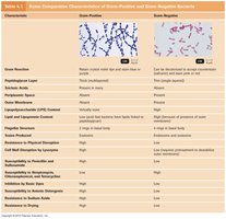
Comparison Table: Gram-Positive vs. Gram-Negative Bacteria
Characteristic | Gram-Positive | Gram-Negative |
|---|---|---|
Peptidoglycan Layer | Thick | Thin |
Teichoic Acids | Present | Absent |
Outer Membrane | Absent | Present |
Endotoxin | Absent | Present (Lipid A) |
Antibiotic Sensitivity | Penicillin | Tetracycline |
Staining | Retains crystal violet | Does not retain crystal violet |
Atypical Cell Walls
Some bacteria possess atypical cell walls, affecting their staining and resistance properties.
Acid-Fast Cell Walls: Contain mycolic acid, characteristic of Mycobacterium and Nocardia.
Mycoplasmas: Lack cell walls, contain sterols in plasma membrane.
Archaea: May lack cell walls or have walls of pseudomurein (lack NAM and D-amino acids).
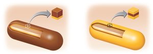
Damage to the Cell Wall
Cell wall integrity is vital for bacterial survival; certain agents can disrupt it, leading to cell lysis.
Lysozyme: Digests peptidoglycan.
Penicillin: Inhibits peptide bridges in peptidoglycan.
Protoplast/Spheroplast: Wall-less cells susceptible to osmotic lysis.
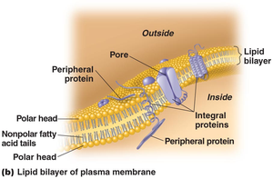
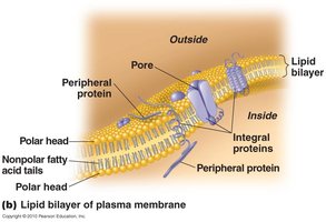
The Plasma Membrane
The plasma membrane is a selectively permeable barrier, crucial for transport, energy production, and cell signaling.
Structure: Phospholipid bilayer with peripheral and integral proteins.
Fluid Mosaic Model: Membrane components move fluidly, allowing dynamic function.
Damage: Alcohols, detergents, and antibiotics can disrupt membrane integrity.
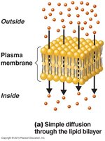
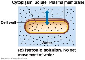
Movement of Materials Across Membranes
Cells transport materials across membranes via several mechanisms, maintaining homeostasis and acquiring nutrients.
Simple Diffusion: Movement from high to low concentration.
Facilitated Diffusion: Solute combines with transporter protein.
Active Transport: Requires transporter protein and ATP.
Osmosis: Movement of water across a selectively permeable membrane.
Endocytosis: Includes phagocytosis (engulfing particles) and pinocytosis (engulfing fluids).
Cytoplasm and Organelles
The cytoplasm contains the cellular machinery for metabolism, growth, and genetic processes.
Cytosol: Fluid portion.
Cytoskeleton: Microfilaments, intermediate filaments, microtubules.
Cytoplasmic Streaming: Movement of cytoplasm throughout cells.
Eukaryotic Organelles
Eukaryotic cells contain specialized organelles for compartmentalized functions.
Nucleus: Contains chromosomes.
Endoplasmic Reticulum (ER): Transport network.
Golgi Complex: Membrane formation and secretion.
Lysosome: Digestive enzymes.
Vacuole: Storage and support.
Mitochondrion: Cellular respiration.
Chloroplast: Photosynthesis.
Peroxisome: Oxidation of fatty acids, destroys H2O2.
Centrosome: Protein fibers and centrioles.
Inclusions and Endospores
Bacteria may contain inclusions for storage and endospores for survival under harsh conditions.
Inclusions: Polysaccharide granules, lipid inclusions, energy reserves.
Endospores: Survival mechanism, resistant to desiccation, heat, chemicals; formed by Bacillus and Clostridium.
Sporulation: Endospore formation; Germination: Return to vegetative state.
Flagella and Cilia in Eukaryotes
Flagella and cilia are motility structures found in eukaryotic cells such as protozoa.
Flagella: Longer, fewer per cell.
Cilia: Shorter, numerous, used for movement and feeding.
Key Terms and Definitions
Peptidoglycan: Major component of bacterial cell wall.
Teichoic Acid: Found in gram-positive cell walls.
Lipopolysaccharide (LPS): Found in gram-negative cell walls, contains endotoxin.
Endospore: Dormant, resistant structure formed by some bacteria.
Binary Fission: Method of prokaryotic cell division.
Important Equations
Osmosis Principle:
Diffusion Rate:
Summary Table: Cell Wall Components
Component | Gram-Positive | Gram-Negative |
|---|---|---|
Peptidoglycan | Thick | Thin |
Teichoic Acid | Present | Absent |
LPS | Absent | Present |
Outer Membrane | Absent | Present |
Additional info: Academic context and expanded explanations were added to ensure completeness and clarity for exam preparation.
