 Back
BackFunctional Anatomy of Prokaryotic and Eukaryotic Cells: Mini-Textbook Study Notes
Study Guide - Smart Notes
 Tailored notes based on your materials, expanded with key definitions, examples, and context.
Functional Anatomy of Prokaryotic and Eukaryotic Cells
Comparing Prokaryotic and Eukaryotic Cells
Microbiology distinguishes between prokaryotic and eukaryotic cells based on structural and functional features. Understanding these differences is fundamental to the study of microbial life.
Prokaryotes: Cells lacking a membrane-bound nucleus. Includes Bacteria and Archaea.
Eukaryotes: Cells with a true, membrane-bound nucleus. Includes Fungi, Protozoa, Algae, and multicellular organisms.
Feature | Prokaryote | Eukaryote |
|---|---|---|
Chromosomes | Usually one circular, not in membrane | Paired, in nuclear membrane |
Histones | Absent | Present |
Organelles | Absent | Present |
Cell Wall | Bacteria: peptidoglycan; Archaea: pseudomurein | Polysaccharide (when present) |
Ribosomes | 70S | 80S (70S in mitochondria) |
Cell Division | Binary fission | Mitosis |
The Size, Shape, and Arrangement of Bacterial Cells
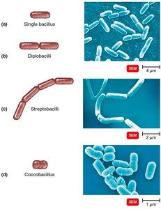
Bacteria exhibit a variety of shapes and arrangements, which are important for identification and classification.
Average size: 0.2–2.0 μm diameter, 2–8 μm length
Monomorphic: Single shape; Pleomorphic: Variable shapes
Basic shapes:
Bacillus (rod-shaped)
Coccus (spherical-shaped)
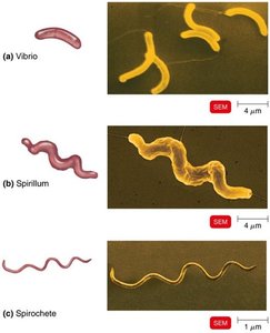
Spiral (includes Vibrio, Spirillum, Spirochete)
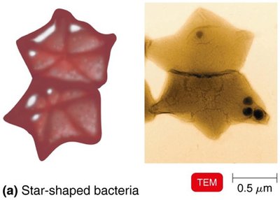
Star-shaped
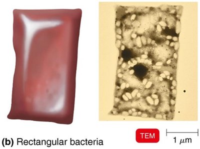
Rectangular
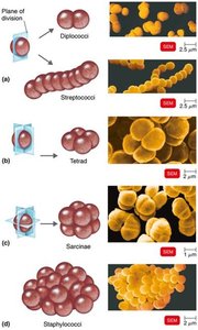
Arrangements:
Pairs: diplococci, diplobacilli
Clusters: staphylococci
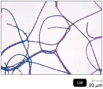
Chains: streptococci, streptobacilli
Groups of four: tetrads
Cubelike groups of eight: sarcinae
Structures External to the Cell Wall
External structures contribute to bacterial survival, pathogenicity, and motility.
Glycocalyx
External to cell wall; viscous and gelatinous
Made of polysaccharide and/or polypeptide
Types: Capsule (organized, firmly attached), Slime layer (unorganized, loose)
Functions:
Contributes to virulence (prevents phagocytosis, aids adherence)
Forms biofilms (protects cells, aids attachment)
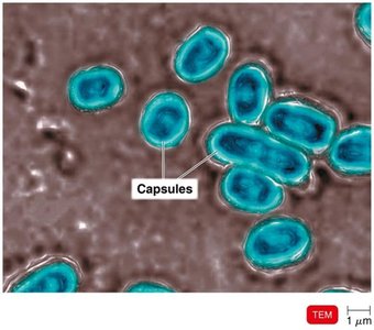
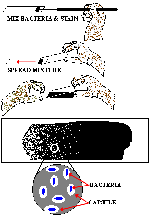
Negative Stain for Capsules
Capsules are visualized by negative staining (e.g., nigrosine, safranine)
Capsules appear as clear halos around colored bacteria against a dark background
Biofilms
Microbial communities encased in a slimy layer
Quorum sensing enables coordinated activity
Biofilms provide protection, facilitate nutrient sharing, and genetic exchange
Significant in human health: increased resistance to antimicrobials, associated with medical device infections
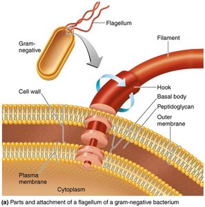
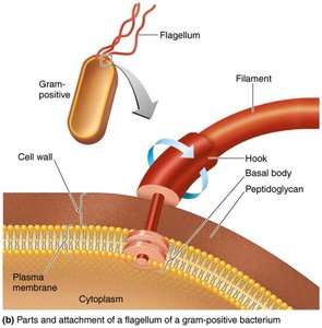
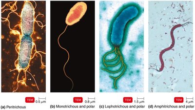
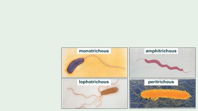
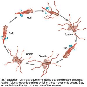
Flagella
Filamentous appendages for motility
Composed of flagellin protein
Three parts: filament, hook, basal body
Arrangement varies: peritrichous, monotrichous, lophotrichous, amphitrichous
Flagella rotate for movement (run/tumble), act as H antigens

Archaella
Motility structures in Archaea
Made of glycoproteins (archaellins), rotate like flagella
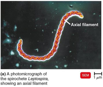
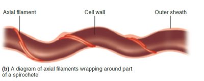
Axial Filaments
Also called endoflagella; found in spirochetes
Anchored at one end; rotation causes corkscrew movement
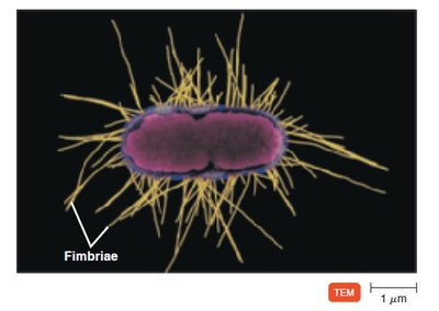
Fimbriae and Pili
Fimbriae: Hairlike appendages for attachment, biofilm formation
Pili: Involved in motility (gliding, twitching) and DNA transfer (conjugation)
The Cell Wall
The bacterial cell wall is a complex structure essential for cell shape, protection, and pathogenicity. Its composition is used to differentiate major groups of bacteria.
Prevents osmotic lysis, protects membrane
Made of peptidoglycan (in bacteria)
Site of action for antibiotics (e.g., penicillin)
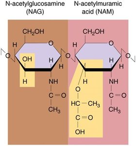
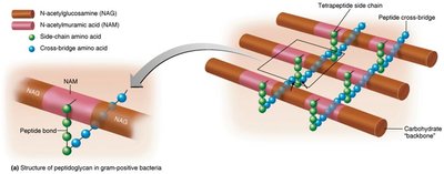
Peptidoglycan Structure
Polymer of repeating disaccharides: N-acetylglucosamine (NAG) and N-acetylmuramic acid (NAM)
Rows linked by polypeptides, forming a lattice
Penicillin interferes with peptide cross-bridges
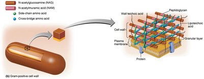
Gram-Positive Cell Walls
Thick peptidoglycan
Teichoic acids (lipoteichoic and wall teichoic acids)
High susceptibility to penicillin, disrupted by lysozyme
Two rings in basal body of flagella
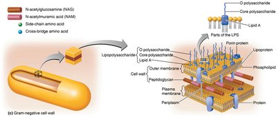
Gram-Negative Cell Walls
Thin peptidoglycan
Outer membrane with lipopolysaccharide (LPS), lipoproteins, phospholipids
Periplasmic space between membranes
Four rings in basal body of flagella
Low susceptibility to penicillin
Cell Walls and Gram Stain Mechanism
Crystal violet-iodine crystals form inside cell
Gram-positive: alcohol dehydrates peptidoglycan, CV-I crystals retained
Gram-negative: alcohol dissolves outer membrane, CV-I washes out, safranin stains cells
Atypical Cell Walls
Acid-fast cell walls: Thick peptidoglycan, waxy mycolic acid, stain with carbolfuchsin
Mycoplasmas: Lack cell walls, sterols in membrane
Archaea: Wall-less or walls of pseudomurein (lack NAM and D-amino acids)
Structures Internal to the Cell Wall
Plasma (Cytoplasmic) Membrane
Phospholipid bilayer enclosing cytoplasm
Peripheral, integral, and transmembrane proteins
Fluid mosaic model: proteins and lipids move freely
Selective permeability, contains enzymes for ATP production
Destruction of Plasma Membrane
Damaged by disinfectants (alcohols, detergents) and antibiotics (polymyxin)
Causes leakage of cell contents
Movement of Materials Across Membranes
Passive processes: No energy required; substances move from high to low concentration
Active processes: Energy required; substances move from low to high concentration
Simple Diffusion
Movement of solute from high to low concentration
Continues until equilibrium is reached
Facilitated Diffusion
Integral membrane proteins act as channels or carriers
Transporter proteins may be nonspecific or specialized
Osmosis
Net movement of water across selectively permeable membrane
Osmotic pressure: pressure needed to stop water movement
Effects of Solutions on Cells
Isotonic: Equal solute concentrations; no net water movement
Hypotonic: Lower solute outside; water enters cell
Hypertonic: Higher solute outside; water leaves cell
Active Transport and Group Translocation
Active transport: requires transporter protein and ATP; moves substances against gradient
Group translocation: requires transporter protein and PEP; substance is chemically altered during transport
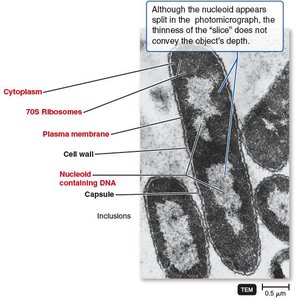
Cytoplasm
Thick, aqueous, elastic substance inside plasma membrane
Contains DNA (nucleoid), ribosomes, inclusions
Cytoskeleton: fibers for cell division, shape, growth, DNA movement
Nucleoid
Bacterial chromosome: circular, double-stranded DNA
Plasmids: small, extrachromosomal DNA circles; carry noncrucial genes (e.g., antibiotic resistance)
Ribosomes
Sites of protein synthesis
Made of protein and ribosomal RNA
70S: 50S (large) + 30S (small) subunits
Target for antibiotics (e.g., streptomycin, gentamicin)
Inclusions
Reserve deposits of nutrients
Types: metachromatic granules (phosphate), polysaccharide granules, lipid inclusions, sulfur granules, carboxysomes, gas vacuoles, magnetosomes
Endospores
Resting cells produced when nutrients are depleted
Resistant to desiccation, heat, chemicals, radiation
Produced by Bacillus and Clostridium
Sporulation: endospore formation; germination: return to vegetative state
Contain dipicolinic acid (DPA) and Ca2+ ions for DNA protection
Summary Table: Gram-Positive vs. Gram-Negative Bacteria
Characteristic | Gram-Positive | Gram-Negative |
|---|---|---|
Peptidoglycan | Thick | Thin |
Teichoic acids | Present | Absent |
Outer membrane | Absent | Present |
Flagella basal body | 2 rings | 4 rings |
Susceptibility to penicillin | High | Low |
Lysozyme sensitivity | High | Low |
Toxins | Exotoxins | Endotoxins & Exotoxins |
Key Equations and Concepts
Peptidoglycan linkage:
Osmosis:
Ribosome assembly:
Additional info:
Biofilms are a major factor in chronic infections and medical device contamination.
Endospores are not reproductive structures; they are survival mechanisms.
Gram stain is a fundamental technique for bacterial classification and diagnosis.
