 Back
BackFunctional Anatomy of Prokaryotic and Eukaryotic Cells: Study Notes
Study Guide - Smart Notes
 Tailored notes based on your materials, expanded with key definitions, examples, and context.
Functional Anatomy of Prokaryotic and Eukaryotic Cells
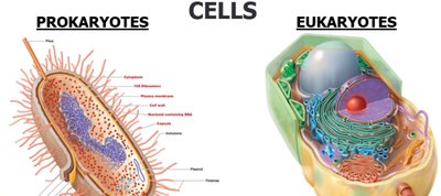
Two Kinds of Cells
Cells are classified into two fundamental types: prokaryotes and eukaryotes. This distinction is foundational in microbiology, as it determines cellular structure, function, and classification of organisms.
Prokaryotes: Include Bacteria and Archaea. No known prokaryotic macroorganisms.
Eukaryotes: Include Animals, Plants, Algae, Fungi, and Protozoa. Both macroorganisms and microorganisms exist in this group.
Prokaryotes | Eukaryotes | |
|---|---|---|
Macroorganisms | None Known | Eukarya: Animals, Plants |
Microorganisms | Archaea, Bacteria | Eukarya: Algae, Fungi, Protozoa |

Cell Structure Comparison
Prokaryotic and eukaryotic cells differ in their internal organization and complexity. Understanding these differences is essential for identifying and studying microorganisms.
Prokaryotes: DNA not enclosed in a nuclear membrane, usually a single circular chromosome, no membrane-bound organelles, complex cell wall if present, divide by binary fission, smaller ribosomes, no cytoskeleton.
Eukaryotes: DNA enclosed in a nuclear membrane, multiple chromosomes, associated with histones and non-histones, membrane-bound organelles (e.g., Golgi complex, mitochondria), simple cell wall if present, divide by mitosis, larger ribosomes, cytoskeleton present.
Feature | Prokaryotes | Eukaryotes |
|---|---|---|
DNA Location | Not enclosed in nuclear membrane | Enclosed in nuclear membrane |
Chromosomes | One circular | Multiple, linear |
Histones | Absent | Present |
Organelles | Absent | Present |
Cell Wall | Complex (if present) | Simple (if present) |
Division | Binary fission | Mitosis |
Size | 0.2–20 μm | 10–100 μm |
Ribosomes | Smaller | Larger |
Reproduction | Asexual (may transfer DNA fragments) | Sexual (meiosis) |
Origin of Terms
The terms prokaryote and eukaryote are derived from Greek:
Prokaryote: "prenucleus"
Eukaryote: "true nucleus"

The Prokaryotes
Prokaryotes include Bacteria and Archaea. They are distinguished by their cellular structure and metabolic diversity.
Some bacteria are photosynthetic (e.g., Cyanobacteria).
Species are differentiated by:
Morphology (shape)
Chemical composition (detected by staining)
Nutritional requirements
Biochemical activities
Source of energy (sunlight or chemicals)
Basic Shapes of Bacteria
Bacteria exhibit a variety of shapes, which are important for identification and classification.
Bacillus: Rod-shaped
Coccus: Spherical
Spiral: Includes Spirillum, Vibrio, and Spirochete

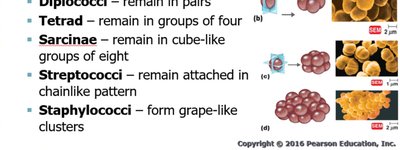
Shapes of Bacterial Cells: Coccus
Cocci are round, spherical, oval, or elongated. Their arrangements are key to identification.
Single
Diplococci: Pairs
Tetrad: Groups of four
Sarcinae: Cube-like groups of eight
Streptococci: Chain-like pattern
Staphylococci: Grape-like clusters
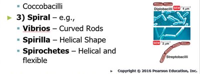
Shapes of Bacterial Cells: Bacillus and Spiral
Bacilli are rod-shaped, and spiral bacteria have distinctive helical forms.
Bacillus: Single, diplobacilli, streptobacilli, coccobacilli
Spiral:
Vibrios: Curved rods
Spirilla: Helical shape
Spirochetes: Helical and flexible
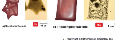
Unusual Bacterial Shapes
Some bacteria exhibit unusual shapes, such as star-shaped (Stella) and square (Haloarcula).
Monomorphic and Pleomorphic Bacteria
Bacterial shape is determined by heredity. Most bacteria are monomorphic (maintain a single shape), but some are pleomorphic (can have more than one genetically controlled shape).
Monomorphic: Environmental factors may change shape, complicating identification.
Pleomorphic: Examples include Rhizobium and Corynebacterium.

Prokaryotic Cell Diagram
The structure of a typical prokaryotic cell includes the plasma membrane, cell wall, capsule, cytoplasm, ribosomes, nucleoid, and flagella.

Structures External to the Cell Wall: Glycocalyx
The glycocalyx is a sugar coat on the surface of many cells, providing protection and aiding in attachment.
Bacterial glycocalyx is viscous (sticky).
Gelatinous polymer of polysaccharide and/or polypeptide.
Made inside and secreted to the cell surface.
Called capsule if organized and firmly attached; slime layer if unorganized and loose.
Structures External to the Cell Wall: Capsule
The capsule is an important virulence factor for pathogenic bacteria.
Provides protection from phagocytosis (e.g., Bacillus anthracis).
Allows attachment to surfaces (e.g., Klebsiella colonization).
Can serve as a source of nutrition when energy stores are low.
Protects against dehydration.
Capsules are antigenic (contain antigens).

Structures External to the Cell Wall: Flagella
Flagella are long filamentous appendages that propel bacteria, contributing to motility and classification.
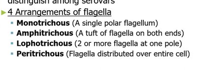
Flagellar protein (H antigen) is used to distinguish among serovars.
Four arrangements:
Monotrichous: Single polar flagellum
Amphitrichous: Tuft of flagella on both ends
Lophotrichous: Two or more flagella at one pole
Peritrichous: Flagella distributed over entire cell

Structure of a Prokaryotic Flagellum
A flagellum consists of three basic parts: filament, hook, and basal body.
Filament: Long, outermost region; composed of flagellin protein arranged in intertwining chains forming a helix around a hollow core.
Hook: Filament attached to it; wider than filament; composed of different protein.
Basal body: Anchors flagellum to cell wall and plasma membrane (not shown in the extracted text but standard academic context).
Additional info: The basal body is a critical component for flagellar function, anchoring the flagellum and enabling rotation for motility.
